Figures & data
Figure 1. Location map of study area (a) showing sites of collect: La Tortuga Island (b) and Turpialito (c). Jacquinia armillaris in their natural habitats TO (d) and TU (e). Detail of leaf blade of TO (f) and TU (g) samples. Scale bars: 1 cm
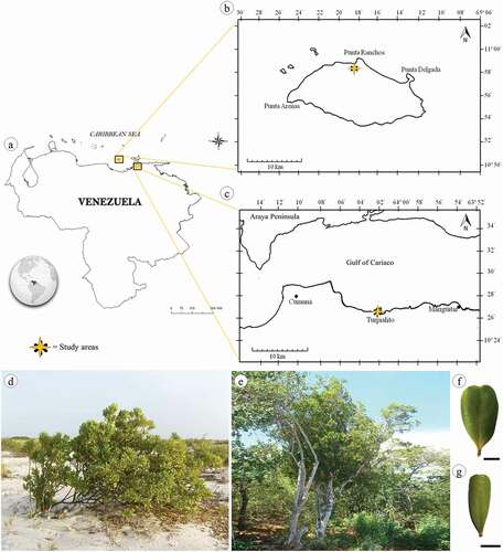
Table 1. Physiochemical characteristics of the soils from the collection sites, La Tortuga (TO) and Turpialito (TU), Venezuela
Table 2. Morphometric characteristics of the leaf blade and petiole of Jacquinia armillaris from La Tortuga (TO) and Turpialito (TU), Venezuela. Different letters indicate differences between localities (t-test at p ≤ 0.05)
Figure 2. Pearson’s correlations coefficients between morphological and anatomical variables of Jacquinia armillaris of La Tortuga and Turpialito, Venezuela. Abbreviations: LBL: leaf blade length; LBW: leaf blade width; LBT: leaf blade thickness; PL: petiole length; PW: petiole width; CT: cuticle thickness; ET: epidermis thickness; LOEC-ad: length of ordinary epidermal cell; WOEC-ad: width of ordinary epidermal cell; NT: number of trichomes; -ad or -ab indicate adaxial or abaxial surface; LGC: length of guard cell; SD: stomatal density; TH: thickness of hypodermis; PPT: palisade parenchyma thickness; SPT: spongy parenchyma thickness. * X indicates correlations with no significance at p ≤ 0.05
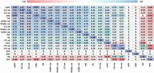
Figure 3. Photographs and microphotographs of cleared leaf showing venation pattern in Jacquinia armillaris from La Tortuga (a) and Turpialito (b). (c,d) Detail of branched secondary and tertiary veins
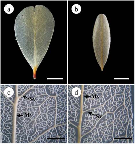
Table 3. Morphometric characteristics of leaf epidermis in Jacquinia armillaris from La Tortuga (TO) and Turpialito (TU), Venezuela. Different letters indicate differences between localities (t-test at p ≤ 0.05). – absence of the characteristic
Figure 4. Epidermis adaxial (a–d) and abaxial (e–h) of Jacquinia armillaris. (a, b, e, f, i, j, k, l): in cross-section. (c, d, g, h): surface view. (a, c, e, g, i, k): of TO samples. (b, d, f, h, j, l): of TU samples. a-b: general aspect of the cuticle and epidermis cells with straight outline of the anticlinal cell walls. (c,d): Epidermis cells, note the depressions. (e,f): Epidermis cells covering by cuticle and stomata guard cells. (g–h): general aspect of cuticle and epidermis straight thickened anticlinal cell walls, note the depressions and stomata. (i–l): Glandular trichomes in depressions in cross-section of epidermis adaxial (i, j) and abaxial (k, l). Abbreviations: cu: cuticle; st: stomata. Scale bars: 50 μm
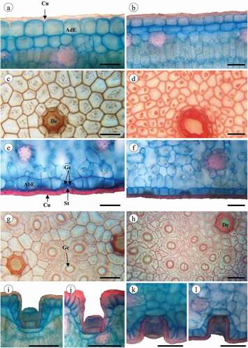
Table 4. Morphometric characteristics of mesophyll in Jacquinia armillaris from La Tortuga (TO) and Turpialito (TU), Venezuela. Different letters indicate differences between localities (t-test at p ≤ 0.05)
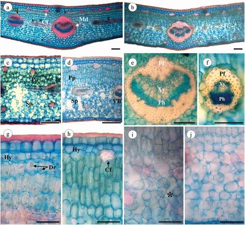
Figure 5. Jacquinia armillaris leaves in cross-section. (a, c, g, i): of TO samples. (b, d, e, f, h, j): of TU samples. (a,b): Leaf section of the primary vein. (c,d): Leaf detail showing palisade and spongy parenchyma. (e,f): primary vein and vascular bundle, xylem and phloem recovery with fibers. (g,h): adaxial region showing the hypodermis and palisade parenchyma cells. (i,-j): Leaf abaxial region, note the spongy parenchyma cells and intercellular spaces (*). Abbreviations: Cf: cortical fibers; Md: midrib; Pp: palisade parenchyma; Sp: spongy parenchyma; VB: vascular bundle; Xy: xylem; Ph: Phloem; Pf: perivascular fibers; Hy: hypodermis; Dr: druses. Scale bars: 100 μm