Figures & data
Figure 1. Principle of the computed micro-tomography system (Soltani et al., Citation2015).
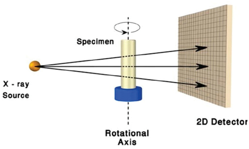
Figure 2. Trabecular bone with metal implants of different diameters: (a) No visible artefacts, (b) With small visible artefacts, (c) With large artefacts, (d) With very large artefacts (arrows indicate artefacts; Grandmougin et al., Citation2020).
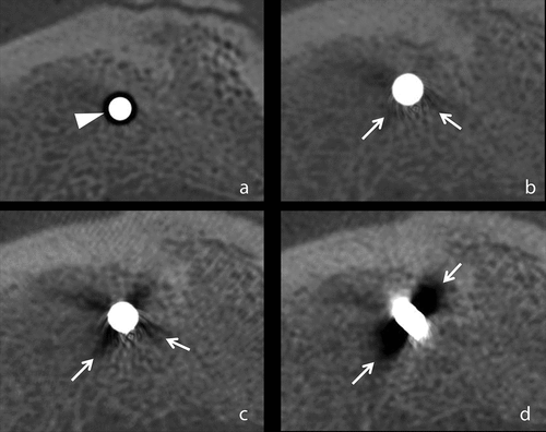
Figure 3. Example of circular artefacts (Kornilov et al., Citation2020).
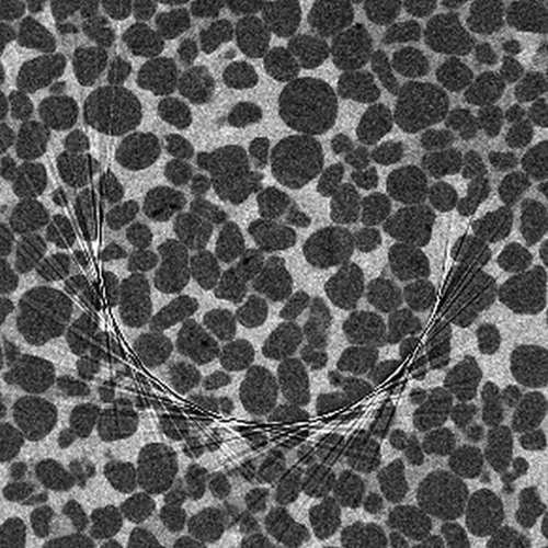
Figure 4. Example of an enlarged image of a “pore space”-”mineral skeleton” rock interface (Perez et al., Citation2022).
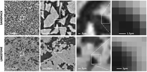
Figure 5. Identification of the pore throat network topology by the “maximum balloon” method: (a) pore bodies filled with spheres of different sizes, (b) schematic of the pore-throat-pore structure (L. Wang et al., Citation2020).
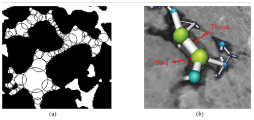
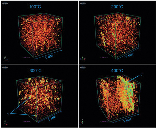
Figure 6. 3D models of the void space structure of heated samples with colour differentiation by the size of the pore channels (cubes with 1 mm edge): 1 – “nodules” of voids, 2 – crack (A. Ponomarev et al., Citation2021).