
Figures & data
Figure 1. Type locality of Mesobiotus sp. nov, Freebird rock pool, Socorro Box Canyon located in near Socorro, New Mexico. Map was made using ArcGIS pro 3.1.1 (ESRI Citation2017) with Chihuahuan Desert boundary from Jornada Basin Spatial Data Laboratory (Citation2006).
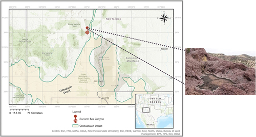
Table I. Sequences used for phylogenetic analysis.
Figure 2. Bayesian phylogenetic reconstruction of the genus Mesobiotus. Numbers above nodes indicate bayesian posterior probability (pp) (shown when pp = 1). Nodes with pp < 0.70 were collapsed. The new described species is highlighted in bold. Scale bar indicates the number of substitutions/site. Outrgoup taxa are not shown. For a complete version of the phylogenetic reconstruction including nodes with pp < 0.70 and outgroups see SM.03.
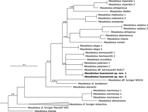
Figure 3. Mesobiotus huecoensis sp. nov. habitus and leg structures under PCM. (a) Holotype habitus. (b) Claws on leg I from paratype on slide SL1. (c) Claws on leg IV from holotype. (d) Granulation on external side of leg I from paratype on slide SL1. (e) Cuticular bulge on the internal side of leg III from paratype on slide SL1. (f) Granulation on legs IV from paratype on slide SL1. Arrowhead: continuous cuticular bar with with shadowed extensions toward the double muscle attachments. Arrow: flexible primary branch on claws IV. Indented arrowhead: horseshoe-shaped structure connects the anterior and posterior lunulae on claws IV. Empty arrowhead: granulation on legs. Empty indented arrowhead: cuticular bulge on internal leg surface. Images a- e were assembled from multiple focus stacks. Scalebars: 10 µm.
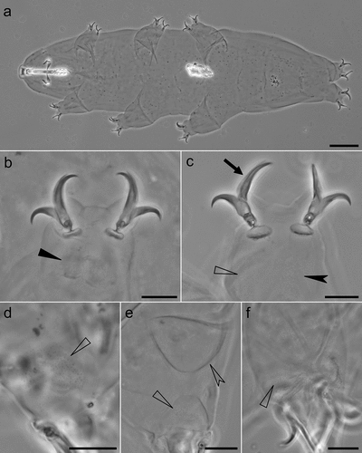
Figure 4. Mesobiotus huecoensis sp. nov. buccopharyngeal apparatus under PCM. (a) Buccopharyngeal apparatus of the holotype. (b) Dorsal view of the placoids row of the holotype. (c) Ventral view of the placoids row of the holotype. (d) Dorsal part of the oral cavity armature (OCA) of the holotype. (e) Dorsal part of the OCA of a paratype on slide SL1. (f) Ventral part of the OCA of the holotype. (g) Ventral part of the OCA of a paratype on slide SL1.Arrowhead: constriction in the third macroplacoid. Arrow: first (anterior) OCA band. Indented arrowhead: second (middle) OCA band. Empty indented arrowhead: third (posterior) OCA band. Empty arrowhead: mucrone behind the median part of the ventral third (posterior) OCA. Image a was assembled from a multiple focus stack. Scalebars: 10 µm.
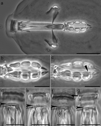
Figure 5. Mesobiotus huecoensis sp. nov. eggs under PCM. (a) General view of the egg showing rounded processes and reticulation on the chorion. (b, c) Sections of egg processes. (d) Egg chorion covered by a reticulum. (e – j) Dorsal view of individual egg processes showing their variability. Arrowheads: pores on the processes. Image a was assembled from a multiple focus stack. Scalebars: (a) 10 µm, (b – j) 5 µm.
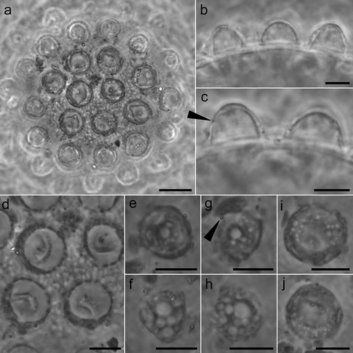
Figure 6. Mesobiotus huecoensis sp. nov. egg under SEM. (a) In toto view. (b) Egg processes showing pores on their lower half. (c) Focus on the reticulated eggshell chorion. Arrowheads: pores on the processes. Indented arrowhead: hole in the chorion reticulum. Scalebars:10 µm.
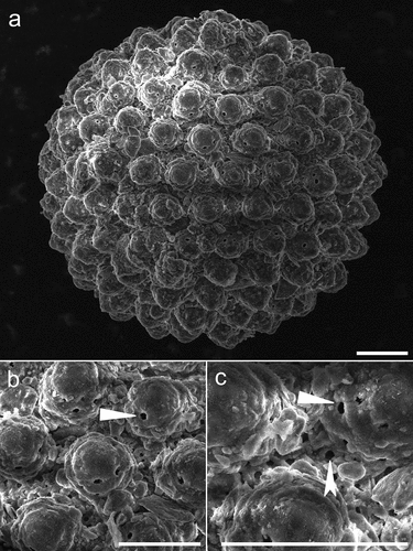
Table II. Measurements [in μm] and pt values of selected morphological structures of individuals of Mesobiotus huecoensis sp. nov.; specimens mounted in Hoyer’s medium.
Table III. Measurements [in μm] of the eggs of Mesobiotus huecoensis sp. nov.; eggs mounted in Hoyer’s medium; process base/height ratio is expressed as percentage.
Table IV. Measurements [in μm] of the sperms of Mesobiotus huecoensis sp. nov. stained with Phalloidin-TRITC and Hoechst 33,342.
Figure 7. Mesobiotus huecoensis sp. nov. male gonad and spermatozoa. (a) Male gonad under PCM stained with orcein showing different maturation stages of the male gamets. (b) Stained spermatozoa under Confocal Microscopy, blue represents stained DNA (nucleus), whereas white represents Phalloidin-stained actin (acrosome, midpiece, tail). Black arrowhead: spermatids. Black indented arrowhead: mature spermatozoa. White arrowhead: acrosome. White indented arrowhead: midpiece. White arrow: tail. Scalebars:10 µm.
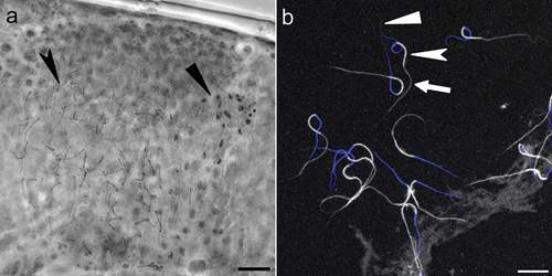
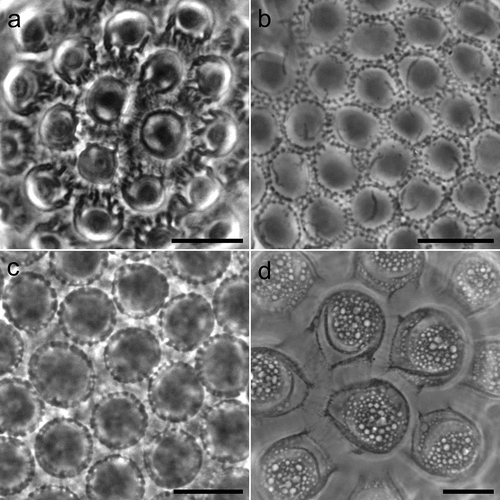
Figure 8. Comparative morphology of Mesobiotus montanus morphogroup species under PCM. (a) M. lusitanicus from type series. (b) M. montanus. (c) M. mottai from type series. (d) M. peterseni. Scale bars:10 µm.
Supplemental Material
Download MS Word (12 KB)Supplemental Material
Download MS Word (282.2 KB)Supplemental Material
Download MS Word (16.9 KB)Supplemental Material
Download MS Word (31.1 KB)Supplemental Material
Download MS Word (12 KB)Supplemental Material
Download MS Excel (11.1 KB)Supplemental Material
Download MS Excel (135.5 KB)Data availability statement
The data that support the findings of this study are openly available in Figshare at DOI:10.6084/m9.figshare.22656586 under CC-BY 4.0 license and in GenBank (see Table 1 for accession numbers).