
Figures & data
Figure 1. Overview of the basic infection cycle depicted by M. anisopliae in invertebrates. The infection process can be divided into: (1) conidia adherence to the host cuticle; (2) conidia germination and development; (3) appressorium formation; (4) cuticle penetration; (5) colonization of hemolymph; (6) extrusion and sporulation.
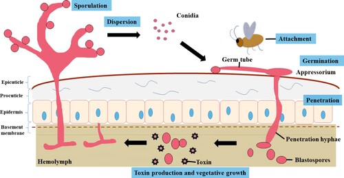
Figure 2. Mycotoxic effects of M. anisopliae. (A) M. anisopliae cultured on Potato Dextrose Agar Medium. At the initial stage, the colonies were white and hairy. In the sporulation stage, there were clumps of green conidia in the middle of the colony. (B) The cadavers of Aedes aegypti infected by M. anisopliae or not. (C) The cadavers of Anopheles stephensi infected by M. anisopliae or not.
Table 1. Agricultural pests and human disease vectors sensitive to M. anisopliae.
Table 2. The major approaches for modifying entomopathogens to enhance the virulence of M. anisopliae.
Table 3. Selected examples of formulations using M. anisopliae in various habitats.
Data availability statement
Data sharing is not applicable to this article as no new data were created or analysed in this study.