275
Views
7
CrossRef citations to date
0
Altmetric
Original

THE EFFECTS OF HEMODILUTION WITH HEMOLINK™ UPON HEMODYNAMICS AND BLOOD FLOW DISTRIBUTION IN ANESTHETIZED DOGS *

, , , , , , , & show all
Pages 137-154 | Published online: 11 Jul 2009

Figures & data

Table 1. Hematocrit and Blood Measurements

Figure 1. Changes in cardiac output (A), mean arterial pressure (B), heart rate (C), stroke volume (D), pulmonary capillary wedge pressure (PCWP) (E), central venous pressure (CVP) (F), systemic vascular resistance (SVR) (G), and pulmonary vascular resistance (PVR) (H), and left (I), and right (J) cardiac work during the 3-hour recovery period following isovolemic exchange–transfusion with Pentaspan (squares); Hemolink-1 (triangles) or Hemolink-2 (upside-down triangles). Values are expressed as a percentage of the baseline value in the same animal prior to the exchange transfusion. Data are expressed as mean±1 SD.

Figure 1. Changes in cardiac output (A), mean arterial pressure (B), heart rate (C), stroke volume (D), pulmonary capillary wedge pressure (PCWP) (E), central venous pressure (CVP) (F), systemic vascular resistance (SVR) (G), and pulmonary vascular resistance (PVR) (H), and left (I), and right (J) cardiac work during the 3-hour recovery period following isovolemic exchange–transfusion with Pentaspan (squares); Hemolink-1 (triangles) or Hemolink-2 (upside-down triangles). Values are expressed as a percentage of the baseline value in the same animal prior to the exchange transfusion. Data are expressed as mean±1 SD.

Table 2. Baseline Hemodynamics in the Pentaspan and Hemolink Groups

Figure 2. Blood flow in the heart (A), brain (B), lung (C), brainstem (D), kidney cortex (E), pancreas (F), hepatic arterial (G), stomach (H), small intestine (I), and large intestine (J), during the 3-hour recovery period following isovolemic exchange- transfusion with Pentaspan® (squares); Hemolink-1 (triangles) or Hemolink 2 (upside-down triangles). Values are expressed as a percentage of the baseline value in the same animal prior to the exchange transfusion. Data are expressed as mean±1 SD. It should be noted that the measured flow to the lung represents the sum of the bronchial arterial blood flow and the very small flow through systemic–venous shunts.

Figure 2. Blood flow in the heart (A), brain (B), lung (C), brainstem (D), kidney cortex (E), pancreas (F), hepatic arterial (G), stomach (H), small intestine (I), and large intestine (J), during the 3-hour recovery period following isovolemic exchange- transfusion with Pentaspan® (squares); Hemolink-1 (triangles) or Hemolink 2 (upside-down triangles). Values are expressed as a percentage of the baseline value in the same animal prior to the exchange transfusion. Data are expressed as mean±1 SD. It should be noted that the measured flow to the lung represents the sum of the bronchial arterial blood flow and the very small flow through systemic–venous shunts.

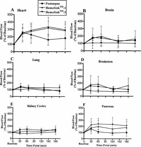
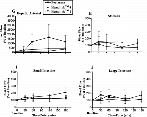
Table 3. Baseline Organ Blood Flows in the Pentaspan and Hemolink Groups

Reprints and Corporate Permissions

Please note: Selecting permissions does not provide access to the full text of the article, please see our help page How do I view content?

To request a reprint or corporate permissions for this article, please click on the relevant link below:

Academic Permissions

Please note: Selecting permissions does not provide access to the full text of the article, please see our help page How do I view content?

Obtain permissions instantly via Rightslink by clicking on the button below:

If you are unable to obtain permissions via Rightslink, please complete and submit this Permissions form. For more information, please visit our Permissions help page.