Figures & data
Figure 1. The variation of proteolytic activity of trout during microwave cooking at different powers (⋄ 20%a, ▪ 40%a, ▴ 60%b). Solid lines represent the model. Powers with different letters are significantly different (p ≤ 0.05).
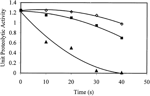
Figure 2. The variation of texture of trout during microwave cooking at different powers (⋄ 20%b, ▪ 40%a,▴ 60%a). Solid lines represent the model. Powers with different letters are significantly different (p ≤ 0.05).
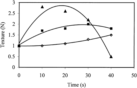
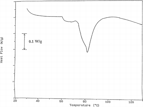
Figure 3. The thermogram obtained for the trout cooked at 40% power for 10 s.
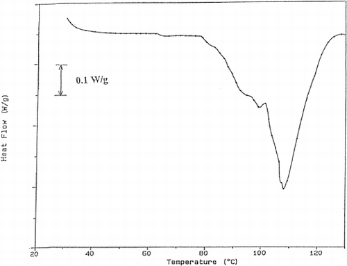
Figure 4. The thermogram obtained for the trout cooked at 40% power for 20 s.
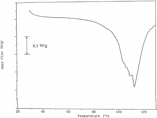
Figure 5. The thermogram obtained for the trout cooked at 40% power for 30 s.
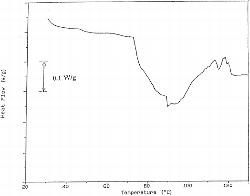
Figure 6. The thermogram obtained for the trout cooked at 40% power for 40 s.