
Figures & data
Table 1. Specific cytogenetic abnormalities in patients who constitute the group of AML abn(3q)
Figure 1. AML with t(3;3)(q21;q26.2). Presence of blasts and dysplastic megakaryocytes in aspirate smear (Wright, x400).
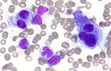
Figure 2. AML with inv(3)(q21q26.2). Bone marrow biopsy showing a marked increase of atypical small mono- and bilobated megakaryocytes (PAS, x400).

Table 2. Main characteristics at diagnosis of the patients in the group of AML with inv(3)(q21q26) or t(3;3)(q21;q26) vs. those with AML carrying other 3q21 or 3q26 abnormality
Table 3. Immunophenotypic features of blasts in AML with inv(3)(q21q26.2) or t(3;3)(q21;q26.2), and those from patients with AML carrying other cytogenetic aberrations involving 3q21 or 3q26