
Figures & data
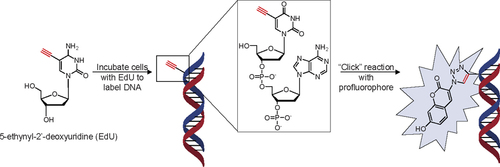
Detection of incorporated nucleotide analog is achieved via reaction of the ethynyl group with a pro-fluorogenic dye, 3-azido-7-hydroxycoumarin. Standard BrdU assays and conventional fluorescent dye labeling of EdU require additional washing steps to remove the unbound fluorescent dyes. In our protocol, these extra wash steps are unnecessary, as the precursor is non-fluorescent until the conjugation between coumarin and EdU is achieved. In brief, the cells are incubated with EdU (10 µM or 50 µM) at 37°C, 5% CO for 30 min to 24 h in complete media (MEM/EBSS supplemented with 10% fetal bovine serum, 1×penicillin, and 1× streptomycin). Then the EdU-labeled cells were fixed with 4% paraformaldehyde for 10 min, washed with PBS, and then reacted with 50 µM dye in buffer solution (100 mM Tris-HCl pH 8.0, 100 mM L-ascorbic acid, 1 mM CuSO4) at room temperature for 1 h.
(A) Fluorescent microscopy images of cells labeled with EdU and reacted with coumarin, and counterstained nuclei with propidium iodide. The cells were seeded on No. 2 coverslips and allowed to adhere to the glass covers overnight. The media was supplemented for the allotted time, after which the cells were fixed with 4% paraformaldehyde for 10 min, washed with PBS, and then reacted with 50 µM dye (100 mM Tris-HCl pH 8.0, 100 mM L-ascorbic acid, 1 mM CuSO4) at room temperature for 1 h. After the reaction, propidium iodide (red) was added to the sample for 10 min. To obtain the best images, the cells on coverslips are rinsed once in water to remove excess salts and placed on glass slides with a PVA/glycerol mounting solution. The samples were examined with an Olympus IX 86 confocal microscope using Semrock DAPI and Cy3 filters. EdU (50 µM) incorporation (blue) gave near complete labeling of cellular DNA after 24 h (images processed with Image Pro Plus V 6.0). (B) Cell labeling efficiency against incubation time and concentration. For short incubation periods, there was no difference in labeling efficiencies for the two different concentrations, but at longer incubation periods, a higher concentration of EdU was necessary to label all of the cells (P < 0.05, n > 200). Cells were counted from four image panels taken randomly with the fluorescent microscope. (C) Three different cell lines were incubated with 50 µM EdU and reacted with azido-coumarin, and counterstained with propidium iodide.


