Figures & data
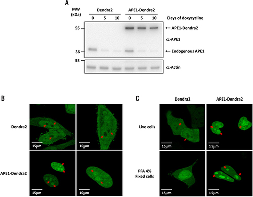
(A) Inducible APE1 siRNA HeLa cell clones were stably transfected with the pDendra2-N empty vector or a vector encoding the APE1-Dendra2 fusion protein. The expression of endogenous and ectopic APE1 was evaluated by Western blotting with total cell extracts before and after expression of the specific APE1 siRNA sequence through doxycycline treatment. Assays were performed by immunoblotting with the specific anti-APE1 antibody or anti-Actin as a loading control. (B) Live confocal analysis of Dendra2 and APE1-Dendra2 clones. Dendra2 protein localizes within cytosol and nuclei but is completely excluded from nucleoli (red arrows). Expression of Dendra2 in fusion with APE1 determines the localization of the recombinant protein within the nuclear compartment and its accumulation in the nucleoli (red arrows). (C) Confocal analysis of HeLa cells expressing Dendra2 and APE1-Dendra2 proteins. After transfection, HeLa cells were analyzed in vivo or fixed with PFA 4% for 20 min. In vivo Dendra2 is excluded from the nucleoli while the fixation procedure generates an artifact showing uniform nuclear staining.
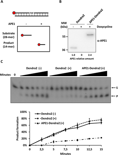
(A) Schematic representation of the enzymatic assay established to measure APE1 endonuclease activity on dsDNA. (B) Western blotting analysis of 5 µg of nuclear cell extracts from Dendra2 and APE1-Dendra2 clones treated (+) or not (-) with doxycycline for 10 days. Relative amounts of APE1 protein are reported at the bottom. (C) APE1 endonuclease activity on abasic dsDNA is rescued by the expression of the APE1-Dendra2 ectopic recombinant protein. The conversion of the radiolabeled THF-containing oligonucleotide substrate (S) to the shorter incised product (P) was evaluated on a denaturing 20% (wt/vol) polyacrylamide gel. A representative image from three independent experiments is shown. Average values of incision percentage with standard deviations of three independent experiments are reported in the graph.
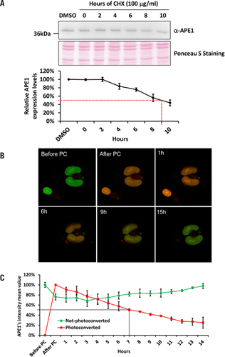
(A) Western blotting analysis of 15 µg of WCE from HeLa cells treated with CHX for the reported amount of time. Densitometric analysis of APE1 relative abundance reported in the graph identify APE1 half-life to be 9 h. Ponceau S staining of the blotted membrane was used as loading control. (B) Representative merged images of APE1-Dendra2 cells analyzed with confocal microscopy before and after Dendra2 photoconversion. (C) Each time point represents the average fluorescence and standard deviation of Dendra2 green (not-photoconverted) and red (photoconverted) calculated on 15 cells before and after UV pulse up to 14 h. The fluorescence half-life of Dendra2 after photoconversion was estimated at 7 h.