Figures & data
Rats received a diffuse axonal brain injury using the Marmarou model then received intranasally applied olfactory mucosa progenitor cells 2 weeks later. At 3 weeks after olfactory mucosa progenitor cell delivery, rats were sacrificed and tissue analyzed.

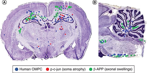
Mapping of OMPC cells and p-c-jun+ and β-APP+ profiles in a coronal (A) and parasagittal section (B) OMPCs were found in injury sites. Injured regions include the cerebral cortex, cerebellum, cerebral peduncles and brain stem tracts. Some of the dots do not appear perfectly circular because of the overlap of dots. In most brains, the number of human OMPCs on each side differed by less than 11%. In this illustrated section, there is more than 11% difference in the number of OMPCs between the two sides and higher indicators of damage (β-APP and p-c-jun) on the side with more OMPCs. A limitation of this study is that labeled cells were not photographed using a confocal microscope to visualize processes of the OMPCs.
OMPC: Olfactory mucosa progenitor cell.