Figures & data
Figure 1 Cardiac position and proximity to the left breast target volume for a patient simulated in both free-breathing and DIBH positions. Position of the heart in free-breathing (A), demonstrating the proximity of the heart to the left breast and nodal target volume. Position of the heart in DIBH (B), demonstrating that the heart is positioned further away from the target volume. When the free-breathing (purple) and DIBH (green) images are fused based on the left breast target volume, the significant difference in cardiac position in the axial (C) and sagittal (D) planes can be appreciated. The position of the heart is contoured in white (solid white, DIBH; dashed white, FB). With DIBH, the heart is displaced inferiorly and medially, further away from the left lateral chest wall.
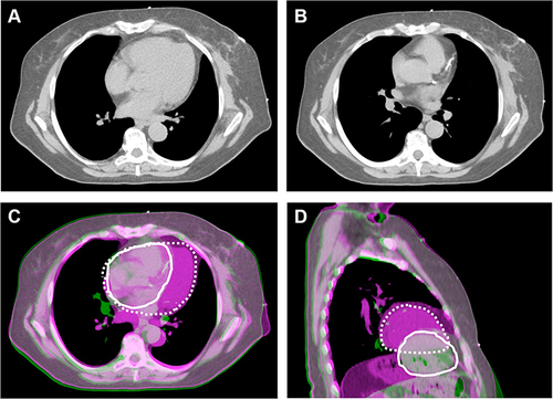
Figure 2 Axial sections with isodose lines for a patient planned for left chestwall and regional nodal treatment (including the internal mammary chain lymph nodes) using four different techniques. (A) Free breathing (FB) 3DCRT, (B) DIBH 3DCRT, (C) FB VMAT, (D) DIBH VMAT.
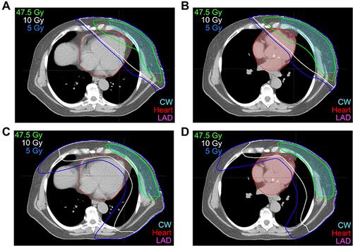
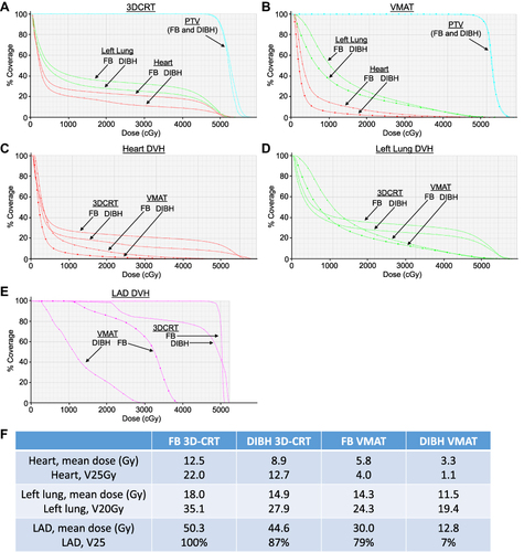
Figure 3 Dosimetric comparison for the heart and left lung, using free-breathing (FB) 3DCRT, DIBH 3DCRT, FB VMAT and DIBH VMAT for the patient shown in . (A) Comparison of dose to PTV (cyan), left lung (green), and heart (red) for FB 3DCRT (closed circles) and DIBH 3DCRT (open circles). (B) Comparison of dose to PTV (cyan), left lung (green), and heart (red) for FB VMAT (triangles) and DIBH VMAT (squares). (C) DVH for the heart with all four planning techniques: FB 3DCRT (closed circles), DIBH 3DCRT (open circles), FB VMAT (triangles), and DIBH VMAT (squares). (D) DVH for the left lung with all four planning techniques: FB 3DCRT (closed circles), DIBH 3DCRT (open circles), FB VMAT (triangles), and DIBH VMAT (squares). (E) DVH for the left anterior descending coronary artery (LAD) with all four planning techniques: FB 3DCRT (closed circles), DIBH 3DCRT (open circles), FB VMAT (squares), and DIBH VMAT (triangles). (F) Comparison of doses to the heart, left lung, and LAD with FB 3DCRT, DIBH 3DCRT, FB VMAT, and DIBH VMAT plans. Note in this patient that DIBH vs FB reduced the percentage of higher doses received by the left lung, heart, and LAD, and that DIBH VMAT showed greater reductions than DIBH 3DCRT and FB VMAT.