Figures & data
Figure 1 (A) Discoid lupus erythematosus (DLE) may present as patchy areas with atrophy and hyperpigmentation. (B) Individual hyperpigmented patches and plaques in discoid lupus erythematosus (DLE) may lack atrophy and simulate pigmented lesions.
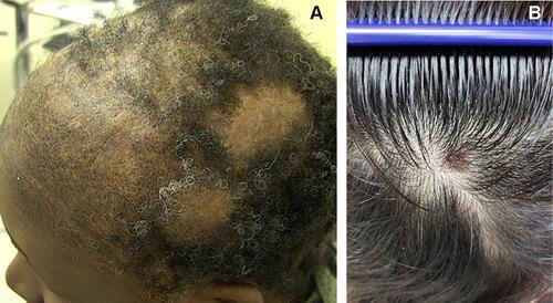
Figure 2 (A and B) Trichoscopy points to keratotic plugs in this case of early discoid lupus erythematosus (DLE) that has been previously diagnosed and treated as alopecia areata (FotoFinder Systems, x40).
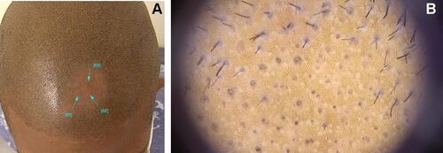
Figure 3 Histologic image of the alopecia areata (AA) subtype of discoid lupus erythematosus (DLE) shows significant interface dermatitis involving the follicular epithelium and increased telogen count (hematoxylin and eosin, x10).
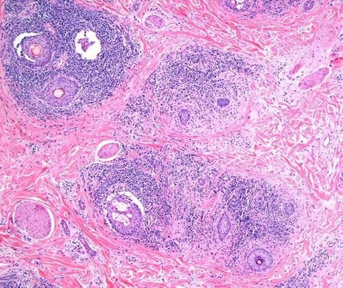
Figure 4 Clinical image demonstrates alopecia in lupus panniculitis of the scalp with notable erythema and lack of keratotic plugs or scale.
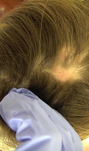
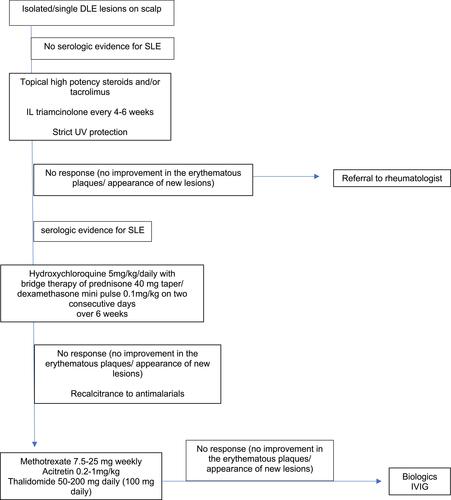
Figure 5 Algorithm describes an approach to treatment of discoid lupus erythematosus (DLE) on the scalp.
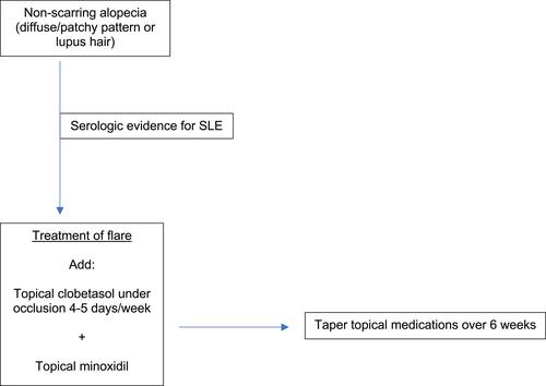
Table 1 Therapies for LE Alopecia
Figure 6 Algorithm describes an approach to treatment of non-scarring alopecia in systemic lupus erythematosus (SLE).