Figures & data
Figure 1 (A) Posterior view of a patient with histopathologically proven diagnosis of AKN in the nape area (red arrow) and FD in the right vertex area (Blue arrow). There is a vast zone of NAS outside of the AKN and FD zones. (B) Same patient showing AKN (nape – red arrow) and FD (right vertex – blue arrow) and a marked parietal NAS area targeted for biopsy. There is a vast zone of NAS outside of the AKN and FD zones.
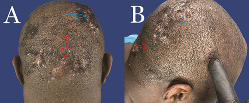
Figure 2 (A) Posterior view of a patient with histopathologically proven diagnosis of AKN in the nape area (red arrow) and FD in the entire vertex area (Blue arrow). There is a vast zone of NAS outside of the AKN and FD zones. (B) Posterior view of a patient with histopathologically proven diagnosis of AKN in the nape area. There is a vast zone of NAS outside of the AKN zone.
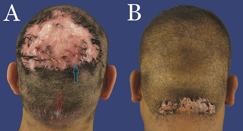
Table 1 Associated Medical Conditions and Past Treatments
Figure 3 (A) Typical trichoscopy finding in the NAS of patients with AKN showing perifollicular scales (Blue arrow) and erythema (Red Arrow), manifesting as hyperpigmentation in a Black African patient. The blue ink mark indicates the biopsy site. (B) Typical trichoscopy finding in the NAS of patients with AKN showing perifollicular scales (Blue arrow) and erythema Red arrow) in a nonblack Hispanic patient. The blue ink mark indicates the biopsy site.
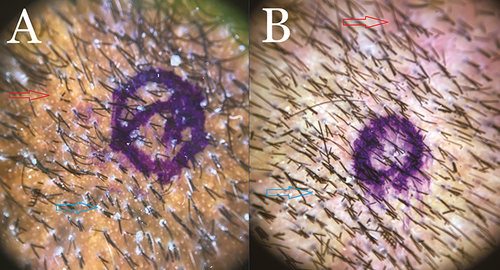
Figure 4 (A) Hematoxylin and eosin stain of the vertical section of an NAS biopsy specimen showing PIILIF. Shows a follicle with lichenoid inflammatory cell infiltrate composed of lymphocytes and plasma cells effacing the dermal-epidermal junction at the level of the infundibulum (Red arrow). There is basal squamatization associated with a Max-Josef space (Green arrow). The interfollicular epidermis is spared of inflammation. Perifollicular and interfollicular fibrosis is present. No sebaceous glands or Vellus or miniaturized hairs are present. Magnification 4x. (B) Hematoxylin and eosin stain of the horizontal section of the same NAS biopsy specimen showing PIILIF. Shows perifollicular inflammatory cell infiltrate composed of lymphocytes and plasma cells effacing the dermal-epidermal junction (Red arrow), basal desquamatization associated with a Max-Josef space (Green arrow), and perifollicular fibrosis (Blue arrows). Magnification 4x.
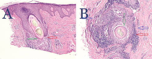
Table 2 Scalp Clinical Characteristics
Table 3 Histological Analysis of the Biopsies for AKN, Healthy Scalp (NAS), and FD
Table 4 Immunohistochemistry (CD4 and CD8 Cells), Mast Cell Activity (CD117 Cells), and Pityrosporum/Demodex Tests
Figure 5 (A) Trichoscopy of normal-appearing beard area of a patient with AKN showing perifollicular and interfollicular erythema Red arrows) and scales (Blue arrows). (B) Hematoxylin and eosin staining of trichoscopy guided-normal appearing in the beard area of a patient with AKN showing PIILIF. A horizontally sectioned follicle with a scant lichenoid inflammatory cell infiltrate composed of lymphocytes and plasma cells, effacing the dermal-epidermal junction associated with perifollicular fibrosis (Blue arrows). Premature desquamation associated with a Max-Josef space (Green arrow) is visible. No Vellus or miniaturized hairs or sebaceous glands are present. Magnification 4x.
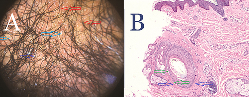
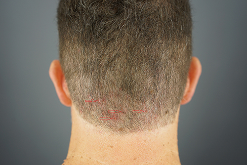
Figure 6 A European-descended White male showing scattered erythematous papules (Red arrows) in the nape area found to be AKN upon histologic analysis.