Figures & data
Table 1 Characteristics of commonly used fillers and their indications
Table 2 Types of dermal filler complication by onset of adverse event
Figure 1 Bruising may be immediate or worsen over 3 days.
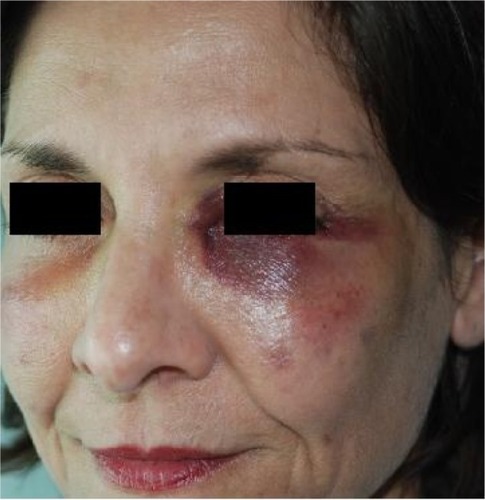
Figure 2 Acute generalized facial edema.
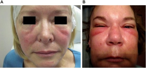
Figure 3 Malar edema.
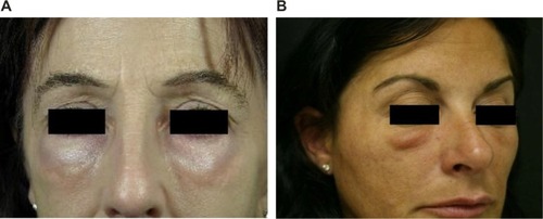
Figure 4 The anatomic basis of malar edema.
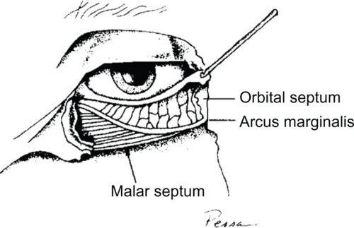
Figure 5 Erythema at site of injection.
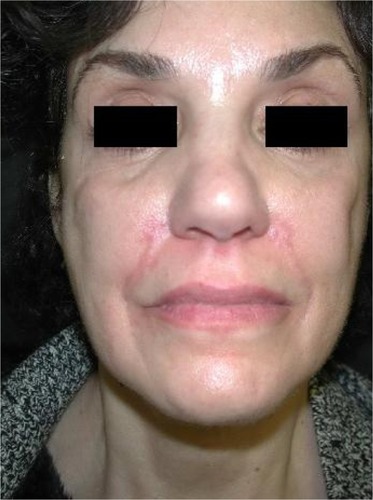
Figure 6 Dyschromia and visible material.
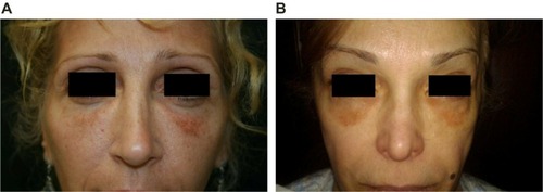
Figure 7 Dermal filler injection leading to herpes virus reactivation.
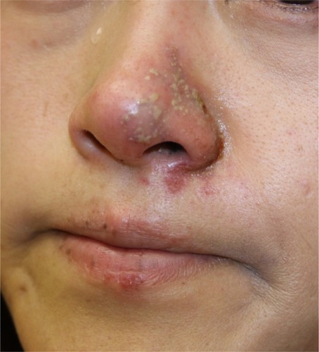
Figure 8 Noninflammatory nodule.
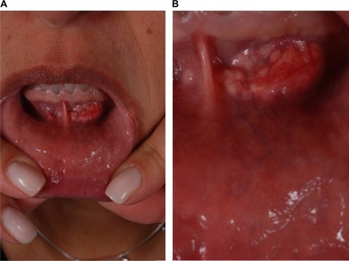
Figure 9 Inflammatory foreign body granuloma before and after treatment with an antibiotic, 5-FU, triamcinolone, and local anesthetic.
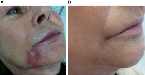
Figure 10 Nodularity that proved to be foreign body granuloma on surgical biopsy.
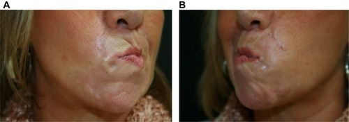
Figure 11 Facial artery anatomy illustrating the most common sites of vascular occlusion.
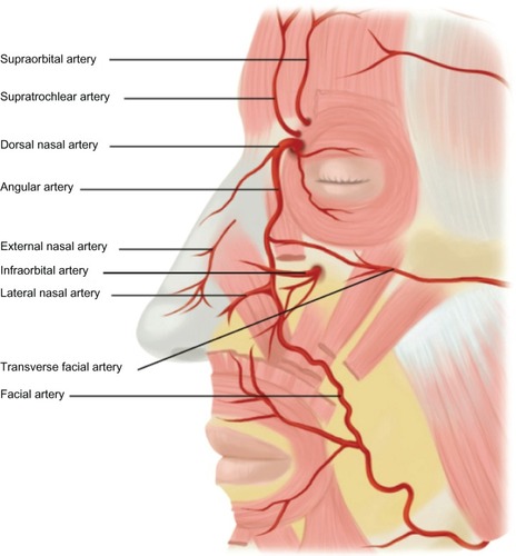
Figure 12 Retinal artery occlusion as a result of calcium hydroxylapatite in the central retinal artery.
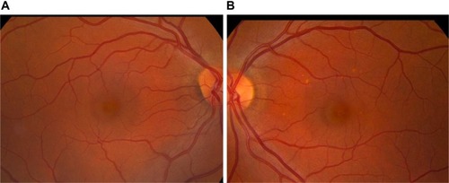
Figure 13 Skin necrosis after dermal filler injection.
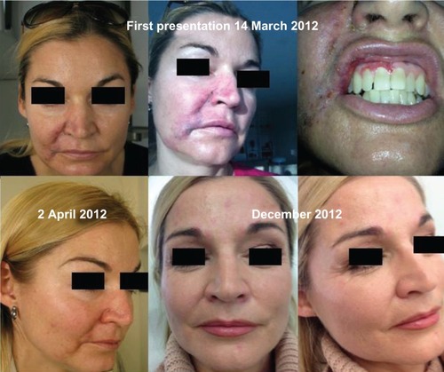
Figure 14 Examples of tissue necrosis after vascular compromise.
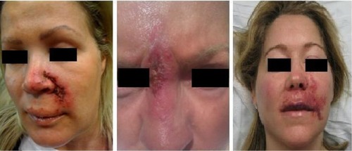
Figure 15 Progression of vascular compromise after an embolic event.
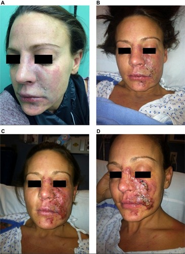
Table S1 Algorithm for the management of antibody-mediated or nonantibody-mediated edema
Table S2 Algorithm for the management of malar edema
Table S3 Algorithm for the management of nodular masses
Table S4 Algorithm for the management of vascular compromise