Figures & data
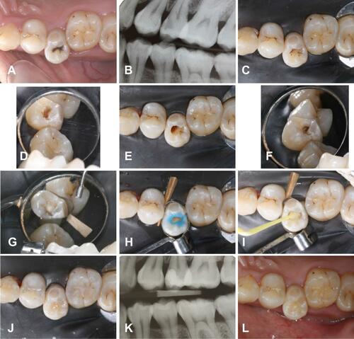
Figure 1 Steps of restoration in Tooth 24. (A) Preoperative image of 24; (B) Preoperative radiographic image of 24; (C) Removal of carious enamel and dentine; (D) Placement of glass ionomer cement liner; (E) Injection of flowable composite into the floor of the cavity; (F) Construction of the distal proximal wall; (G) Removal of the sectional matrix; (H) Filling the cavity using the incremental technique; (I) Reconstruction of the morphology of 24; (J) Sealing the micro-cavitated occlusal fissure caries with a fissure sealant; (K) Postoperative bitewing radiograph; (L) Postoperative image taken three months after the restoration.
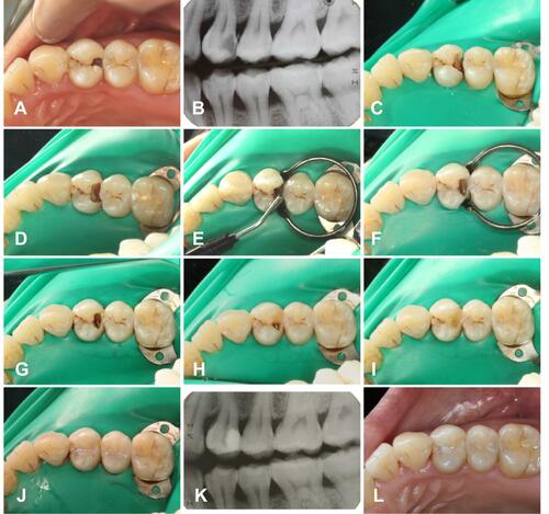
Figure 2 Steps of tunnel restoration in Tooth 15. (A) Occlusal view of 15 before operation; (B) Right bitewing radiography; (C) Occlusal view of the access to the mesial proximal caries; (D) Lateral view of the access to the mesial proximal caries; (E) Occlusal view of the cavity after caries removal; (F) Lateral view of the cavity after caries removal; (G) Restoration of the proximal wall of the cavity; (H) Selective acid-etching on enamel; (I) Bonding agents were applied to the cavity; (J) Restoration of the occlusal cavity; (K) Postoperative bitewing radiograph; (L) Occlusal view after three months.