Figures & data
Table 1 The Prevalence of MB2 Canals According to Gender
Table 2 The Prevalence of MB2 Canals According to Age
Figure 1 Representative CBCT images showing mesiobuccal root canal configurations. Arrows represent position of the mesiobuccal canals in axial sections (coronal, middle, apical thirds), sagittal and coronal sections (left to right). (A) Vertucci type I. (B) Vertucci type II. (C) Vertucci type IV.
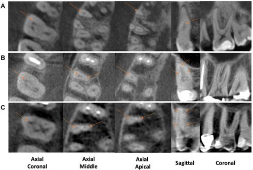
Table 3 The Prevalence of MB2 Canal According to the Location of Joining in the MB Root
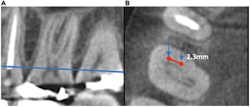
Figure 2 Representative CBCT images showing mesiobuccal root canals and inter-orifice distance at the pulpal floor level. (A) Sagittal section with a blue line representing the level of the pulpal floor. (B) Arrows representing the position of the mesiobuccal canals in an axial section at the level of the pulpal floor. Red line represents the inter-orifice distance measured at the pulpal floor level.