Figures & data
Figure 1 The process of mono-ADP-ribosylation (MARylation). ADP-ribosylation is reversible; it is catalyzed by ADP-ribosyltransferases (ARTs) and removed by ADP-ribosylglycohydrolases.
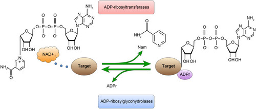
Table 1 Different Degrees of Mono-ADP-Ribose Binding Reagent Staining in Colorectal Adenocarcinoma and Normal Colorectal Tissue
Table 2 Mono-ADP-Ribose Binding Reagent in the Nucleus of Colorectal Adenocarcinoma and Normal Colorectal Tissue
Table 3 Mono-ADP-Ribose Binding Reagent in the Cytoplasm of Colorectal Adenocarcinoma and Normal Colorectal Tissue
Figure 2 Staining of mono-ADP-ribose binding reagent in colorectal adenocarcinoma tissue samples. (A) Strongly positive staining (×400). (B) Slight positive staining (×400). Scale bar=50μm.
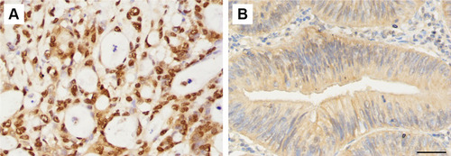
Figure 3 The level of mono-ADP-ribose binding reagent in colorectal adenocarcinoma and normal colorectal tissues. Positive staining of mono-ADP-ribose binding reagent in colorectal adenocarcinoma (A–C) (×4), (D–F) (×400). Positive staining of mono-ADP-ribose binding reagent in normal colorectal tissue (G–I) (×400). Scale bar=50μm.
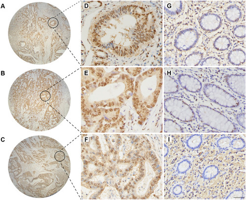
Table 4 Comparison of Clinicopathological Characteristics in 64 Cases with Colorectal Adenocarcinoma
Table 5 The Difference Analysis of Invasive Depth and the Level of Mono-ADP-Ribose Binding Reagent in Cytoplasm
Table 6 The Difference Analysis of Histological Classification and the Level of Mono-ADP-Ribose Binding Reagent in Nucleus
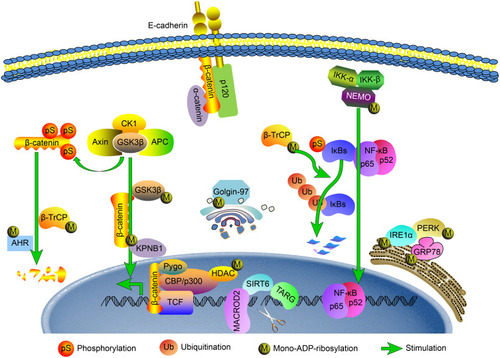
Figure 4 Schematic diagram of how MARylation is predicted to be involved in the regulation of colorectal cancer. The possible mechanisms of MARylation in colorectal cancer are described with regard to endoplasmic reticulum stress, DNA damage repair, EMT, β-catenin activity and the NF-κB signaling pathway.