Figures & data
Figure 1 Large carcinoma of the cecum (red circle) as displayed on double contrast barium enema image. Lesion presence is inferred indirectly as a filling defect of the cecal lumen with irregular mucosal lining.
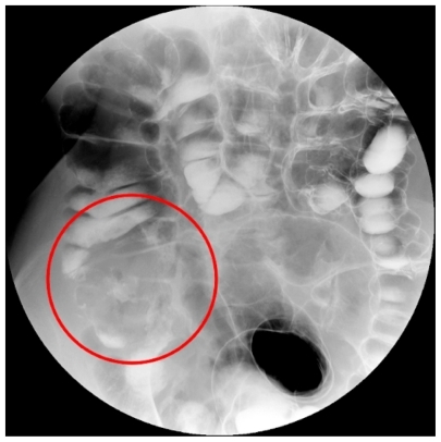
Figure 2 Computed tomography colonography image in the supine a) and prone b) position. In the supine position the collapsed sigmoid colon may mimic cancer, while on the prone position the bowel walls and the lumen are shown to be normal (red arrows).
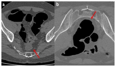
Figure 3 a) Annular stenosing cancer of the right colonic flexure (red circle) using a low dose computed tomography colonography (CTC) protocol. b, c) CTC can be performed with a regular dose protocol for detection of extracolonic disease, such as lung (b: white circle) and liver metastases (c: red arrows) in a patient with locally advanced colorectal cancer (red asterisk).
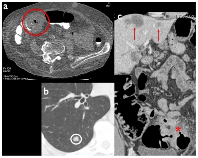
Figure 4 Sessile polyp of the ascending colon: a) native axial image (red arrow), b) virtual endoscopic view (red asterisk).
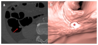
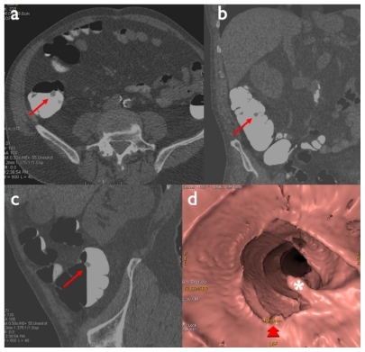
Figure 5 Pedunculated polyp of the ascending colon: a) native axial image, b) coronal reformation, c) sagittal reformation (red arrows), d) virtual endoscopic view (white asterisk).