Figures & data
Table 1 Clinical characteristics of nine smokers with COPD, eight asymptomatic smokers, and 13 never-smokers
Table 2 Cell characteristics of nine smokers with COPD, eight asymptomatic smokers, and 13 never-smokers
Figure 1 IL-16 protein (A) and IL-1β protein (B) in plasma.
Abbreviations: AS, asymptomatic smokers; IL, interleukin; NS, never-smokers.
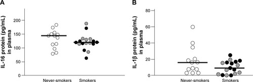
Table 3 Flow cytometry analysis of intracellular IL-16 protein plus the relative and total number of NK cells, CD8+ T-cells, CD4+ T-cells, B-cells, and monocytes as well as rMFI analyzed by flow cytometry
Figure 2 The relative number of IL-16+ NK cells in blood (A). AS are indicated by [Image], NS are indicated by ◯, and smokers with COPD by ●. Data is presented as individual and median values (bold lines). The Mann–Whitney U-test was performed (P<0.05.) A representative flow cytometry analysis was conducted for IL-16+ CD3− CD16+ CD56+ cells (B).
Abbreviations: AS, asymptomatic smokers; COPD, chronic obstructive pulmonary disease; IL, interleukin; IL-16 IC, IL-16 isotype control; NS, never-smokers.
![Figure 2 The relative number of IL-16+ NK cells in blood (A). AS are indicated by [Image], NS are indicated by ◯, and smokers with COPD by ●. Data is presented as individual and median values (bold lines). The Mann–Whitney U-test was performed (P<0.05.) A representative flow cytometry analysis was conducted for IL-16+ CD3− CD16+ CD56+ cells (B).](/cms/asset/37a7c133-dc7c-4143-977e-db9510cb6e15/dcop_a_103758_f0002_c.jpg)
![Figure 2 The relative number of IL-16+ NK cells in blood (A). AS are indicated by [Image], NS are indicated by ◯, and smokers with COPD by ●. Data is presented as individual and median values (bold lines). The Mann–Whitney U-test was performed (P<0.05.) A representative flow cytometry analysis was conducted for IL-16+ CD3− CD16+ CD56+ cells (B).](/cms/asset/a0a2e9ef-ce46-4797-bd0d-30d78097d790/dcop_a_103758_f0002b_c.jpg)
Figure 3 Correlations between, on one hand, the relative (A) and the total (B) numbers of NK cells and CD4+ T-cells (C) in blood from the pooled group of smokers (COPD + AS) and, on the other, tobacco load in pack-years (Spearman rank correlation: P<0.05, ρ: −0.56 [A and B] and −0.62 [C]).
![Figure 3 Correlations between, on one hand, the relative (A) and the total (B) numbers of NK cells and CD4+ T-cells (C) in blood from the pooled group of smokers (COPD + AS) and, on the other, tobacco load in pack-years (Spearman rank correlation: P<0.05, ρ: −0.56 [A and B] and −0.62 [C]).](/cms/asset/9447be52-3396-4715-b5d9-be8f102285ca/dcop_a_103758_f0003_b.jpg)
Figure 4 Total number of activated (CD69+) CD8+ T-cells in blood for never-smokers, asymptomatic smokers and smokers with COPD.
Abbreviation: COPD, chronic obstructive pulmonary disease.
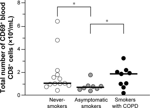
Figure 5 Concentrations of extracellular IL-16 protein measured by ELISA in conditioned medium from human blood NK cell cultured for 20 hours in vitro, exposed to either water-soluble tobacco smoke components (CSE) or vehicle solution (VEH) and treated with glutathione (10−2 M).
Abbreviations: CSE, cigarette smoke extract; IL, interleukin; VEH, vehicle.
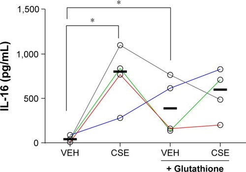
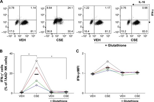
Figure 6 Flow cytometry analysis of human NK cells cultured for 20 hours in vitro exposed to either water-soluble tobacco smoke components (CSE) or vehicle solution (VEH) and treated with glutathione (10−2 M).
Abbreviations: CSE, cigarette smoke extract; IL, interleukin; rMFI, relative mean fluorescence index; VEH, vehicle.
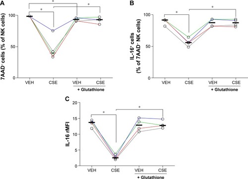
Figure 7 Result of flow cytometry analysis of human NK cells cultured for 20 hours in vitro, exposed to either water-soluble tobacco smoke components (CSE) or vehicle solution (VEH) and treated with glutathione (10−2 M).
Abbreviations: CSE, cigarette smoke extract; rMFI, relative mean fluorescence index; VEH, vehicle.