Figures & data
Figure 1 Three different pathways for collateral ventilation.
Notes: Interalveolar ventilation through the pores of Kohn. Bronchiole–alveolar ventilation through channels of Lambert. Interbronchiolar ventilation through channels of Martin.
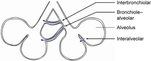
Figure 2 Collateral ventilation.
Notes: Schematic figure illustrating the role of intra- and interlobar collateral ventilation, and the implications for bronchoscopic lung volume reduction using one-way endobronchial valves. The interlobar collateral ventilation from the left upper to the left lower lobe, impedes the desired atelectasis of the lower lobe. Because all segments of the left lower lobe have endobronchial valves, intralobar collateral ventilation does not influence the development of atelectasis.
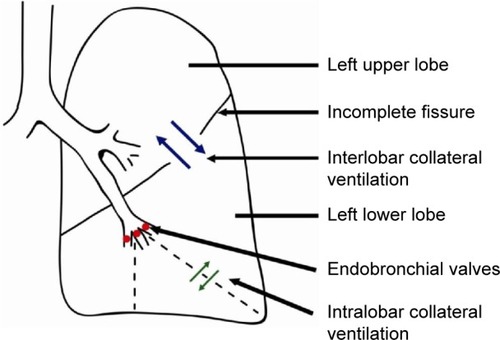
Table 1 Prevalence of incomplete fissures
Figure 3 Chartis measurement.
Notes: (A) The Chartis balloon at the distal tip of the catheter. (B) Bronchoscopic view of the Chartis balloon blocking the entrance to the right lower lobe to measure collateral ventilation to this lobe. (C) Example of a negative Chartis measurement with absence of collateral ventilation, measured in spontaneous breathing patient. The orange pattern shows the expired flow (mL/min). The decrease of the flow pattern indicates there is no collateral flow. The blue pattern shows the negative intrapleural pressure (cmH2O) and indicates the quality of the occlusion by the balloon. (D) Example of a positive Chartis measurement with collateral ventilation, as there is no decline in the expired flow. (E) Example of a negative Chartis measurement with absence of collateral ventilation, measured in a sedated patient with positive pressure ventilation. Therefore, only the decreasing flow pattern is shown, indicating there is no collateral flow.
Abbreviations: F, flow; P, pressure.
Abbreviations: F, flow; P, pressure.
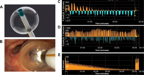
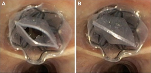
Figure 4 Implanted one-way endobronchial valve.
Notes: (A) Open valve, allowing trapped air and fluids to escape. (B) Closed valve, no air or fluids can enter the valve.