Figures & data
Table 1 Patient characteristics
Figure 1 Multidetector computed tomography image.
Note: Lobar atelectasis of the right lower lobe following valve implantation.
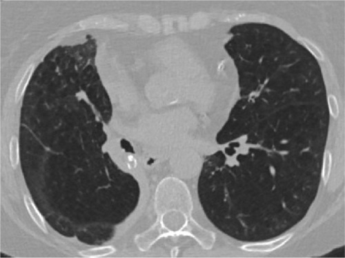
Figure 2 Multidetector computed tomography image.
Notes: Pneumothorax with subsequent severe bilateral subcutaneous emphysema 3 days following valve placement in the left lower lobe. The lung is reinflated after chest tube insertion.
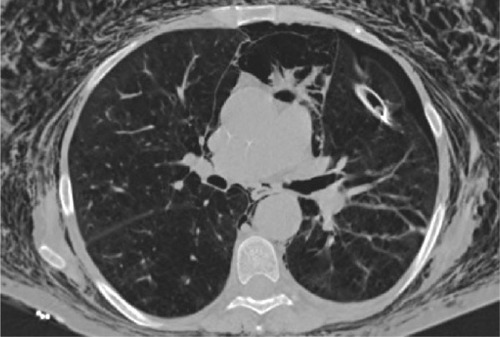
Table 2 Clinical outcome of patients who developed pneumothorax
Table 3 Predictors for pneumothorax following valve therapy
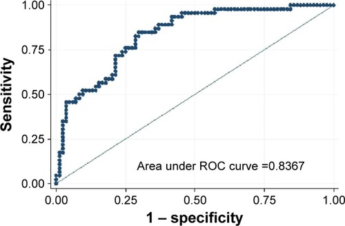
Figure 3 ROC curve.
Note: Prediction of pneumothorax following endoscopic valve therapy using eight CT and clinical parameters with an AUC of 0.8367.
Abbreviations: CT, computed tomography; AUC, area under the curve; ROC, receiver operating characteristic.
Abbreviations: CT, computed tomography; AUC, area under the curve; ROC, receiver operating characteristic.