Figures & data
Table 1 Incidence of thin-section CT findings in nonsmokers and smokers (172 cases and 1,032 lobes)
Figure 1 Incidence of thin-section computed tomography findings in nonsmokers and smokers (172 cases and 1,032 lobes).
Abbreviations: LAA, low attenuation area; CCVW, clustered cysts with visible walls; GGAR, ground-glass attenuation with/without reticulation; DICN, diffuse ill-defined centrilobular nodules.
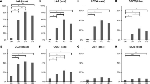
Table 2 Incidence of thin-section CT findings among lobes and zones
Figure 2 Incidence of thin-section computed tomography findings among lobes and zones.
Abbreviations: LAA, low attenuation area; CCVW, clustered cysts with visible walls; GGAR, ground-glass attenuation with/without reticulation; DICN, diffuse ill-defined centrilobular nodules.
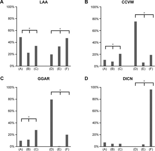
Table 3 Correlation of thin-section CT findings with histological findings in 172 resected lobes
Table 4 Correlation of additional thin-section CT findings of CCVW with histological findings in 30 resected lobes
Figure 3 Severe SRIF with emphysema pattern on thin-section CT images and histological findings.
Notes: (A and B) Thin-section CT images showing clustered cysts of markedly irregular size and shape accompanied by ground-glass attenuation with reticular structures in the surrounding area. (C) Low-power photograph of a histological section (hematoxylin–eosin stain) reveals irregularly shaped emphysematous spaces (asterisk) with collagenous fibrotic walls. Many of the walls are truncated. Patchy fibrosis is observed in the intervening lung parenchyma and corresponds to ground-glass attenuation with reticular structures on thin-section CT. Irregular cysts with thickened walls tend to be present a little apart from the pleura with less-involved subpleural lung parenchyma. (D) On this high-power photograph of a histological section, fibrosis consists of hyalinized paucicellular fibrosis (arrow) corresponding to SRIF.
Abbreviations: SRIF, smoking-related interstitial fibrosis; CT, computed tomography.
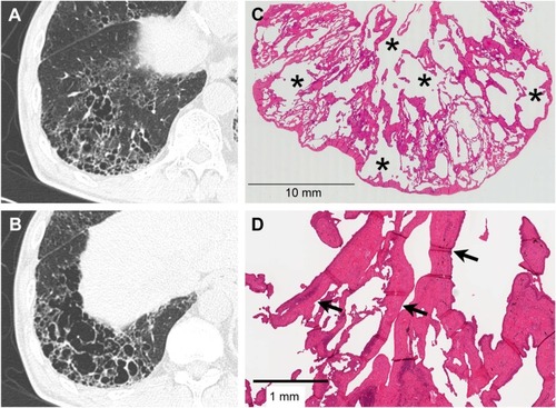
Figure 4 Mild SRIF with emphysema pattern on thin-section CT images and histological findings.
Notes: (A and B) Thin-section CT images showing clustered cysts with visible walls in the peripheral zone with less involvement of the subpleural parenchyma. The size and shape of the cysts vary. Note ground-glass attenuation with reticular or branching structures in the surrounding area. (C) This low-power photograph of the histological section (hematoxylin–eosin stain) reveals irregularly shaped emphysematous spaces (asterisk) with thickened fibrotic walls corresponding to clustered cysts with visible walls on the thin-section CT image. There is patchy fibrosis at peribronchiolar and subpleural sites corresponding to ground-glass attenuation with reticular or branching structures in the surrounding area. Irregular cysts with thickened walls are present apart from the pleura with less-involved subpleural lung parenchyma. (D) On this high-power photograph of a histological section, fibrosis of this pattern consists of hyalinized paucicellular fibrosis (arrow) or SRIF.
Abbreviations: SRIF, smoking-related interstitial fibrosis; CT, computed tomography.
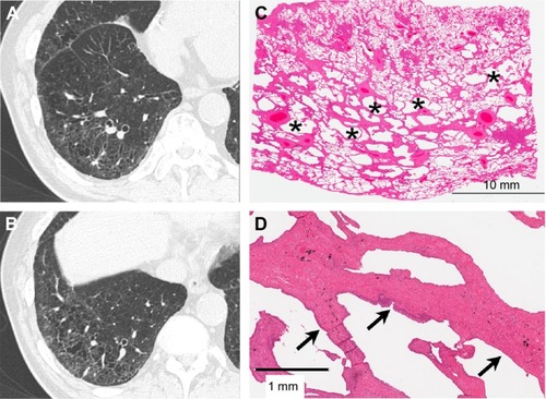
Figure 5 UIP pattern on thin-section CT images and histological findings.
Notes: (A and B) Thin-section CT images showing clustered cysts of similar size and shape. Note surrounding ground-glass attenuation with reticular opacity. (C) This low-power photograph of the histological section (hematoxylin–eosin stain) reveals macroscopic honeycombing (asterisk) corresponding to honeycombing on thin-section CT images and patchy fibrosis with architectural distortion and microscopic honeycombing reflecting ground-glass attenuation with reticular opacity. (D) Note dense fibrosis and fibroblastic foci (arrow) on high-power photographs of the histological section. (E) Masson’s trichrome stain reveals architecture destruction with scattered fibroblastic foci (arrow).
Abbreviations: UIP, usual interstitial pneumonia; CT, computed tomography.
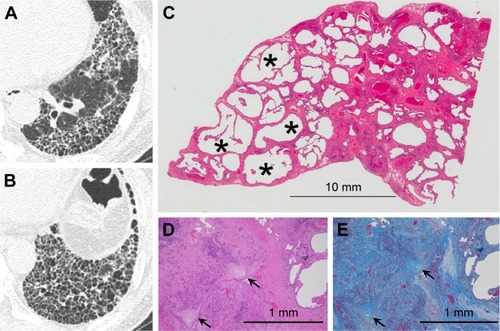
Figure 6 Combined SRIF and UIP patterns on thin-section CT images and histological findings.
Notes: (A) Thin-section CT images showing foci of clustered cysts of irregular size and shape and surrounding ground-glass attenuation with reticular or branching structures. (B) This low-power photograph of the histological section (hematoxylin–eosin stain) reveals areas with irregularly shaped emphysematous spaces (asterisk) and hyalinized fibrotic walls (arrow). (C) High-power photograph of a histological section. Note dense fibrosis and fibroblastic foci (arrow) in the same histological specimen. (D) Masson’s trichrome stain reveals architecture destruction with irregularly distributed dense fibrosis. The fibroblastic focus (arrow) is seen inside the fibrosis.
Abbreviations: SRIF, smoking-related interstitial fibrosis; UIP, usual interstitial pneumonia; CT, computed tomography.
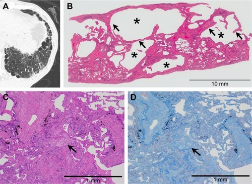
Figure 7 Combined SRIF and UIP patterns on thin-section CT images.
Abbreviations: SRIF, smoking-related interstitial fibrosis; UIP, usual interstitial pneumonia; CT, computed tomography.
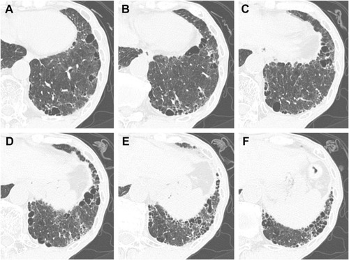
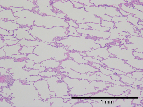
Figure S1 High-power photograph of a normal lung.
Notes: The parenchyma of normal lung shows little fibrotic change compared with SRIF. The architectural destruction and emphysematous change are absent in normal lung.
Abbreviation: SRIF, smoking-related interstitial fibrosis.