Figures & data
Table 1 Clinical data of study participants as a function of presence and severity of COPD
Table 2 Results obtained by screening for sleep-disordered breathing as a function of presence and severity of COPD
Figure 1 ODI, stratified by the presence of overlapping OSA in COPD patients.
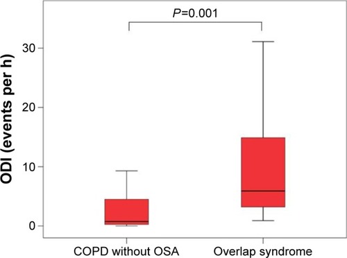
Table 3 Comparison of laboratory, electrocardiographic, as well as conventional and speckle tracking-based echocardiographic data according to presence and severity of COPD
Figure 2 Left ventricular longitudinal strain analysis, visualized by two-dimensional speckle tracking echocardiography.
Notes: Deformation imaging of the left ventricle was performed in both COPD patients (A) and controls (B), which showed significant differences in global and apical longitudinal deformation properties.
Abbreviations: Long, longitudinal; Max, maximum; Seg, segments; PK, peak; TPk, time to peak; GLS, global longitudinal strain; endo, endocardial.
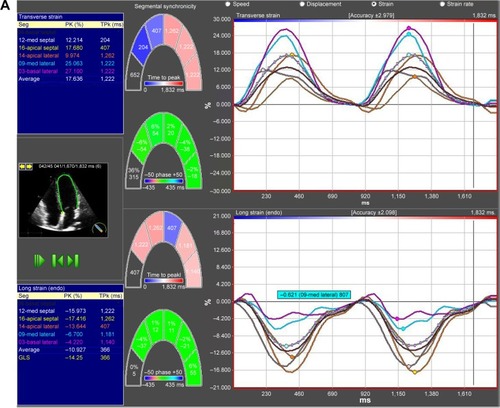
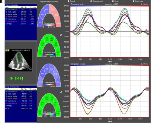
Figure 3 Intercohortal comparison between COPD patients and control smokers for global (A) and apical septal and (B) longitudinal left ventricular strain.
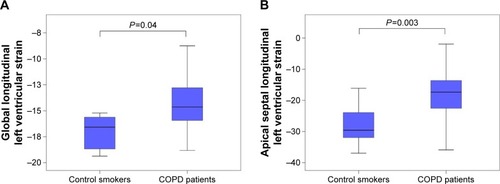
Figure 4 Diagnostic accuracy of apical septal longitudinal LV strain for identification of patients with COPD.
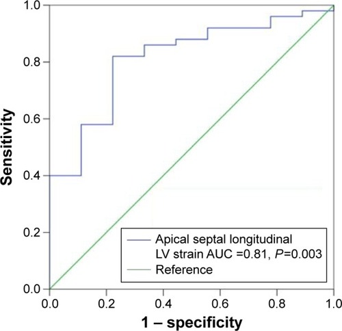
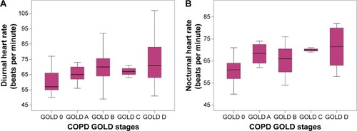
Figure 5 Both diurnal heart rate (A), measured by 12-lead electrocardiogram, and nocturnal heart rate (B), recorded by somnological screening, offer significant increase over COPD GOLD stages (P=0.01 and P=0.001, respectively).
Abbreviation: GOLD, Global initiative for Chronic Obstructive Lung Disease.