Figures & data
Table 1 Demographic characteristics of patients with COPD and healthy controls
Table 2 Regional information of changed ALFF in patients with COPD
Figure 1 Changes of ALFF in patients with COPD compared with controls.
Notes: (A) Uncontrolling for SaO2, (B) controlling for SaO2. P<0.05 (corrected). Red to yellow indicates an increase, blue indicates a decrease.
Abbreviations: ALFF, amplitude of low-frequency fluctuation; COPD, chronic obstructive pulmonary disease; L, left; R, right.
Abbreviations: ALFF, amplitude of low-frequency fluctuation; COPD, chronic obstructive pulmonary disease; L, left; R, right.
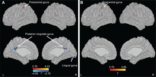
Figure 2 Regional ALFF in the brain of patients with COPD compared with controls by ROI analysis.
Notes: *P<0.05; ***P<0.001. Error bars represent standard deviation.
Abbreviations: ALFF, amplitude of low-frequency fluctuation; COPD, chronic obstructive pulmonary disease; PCC, posterior cingulate cortex; ROI, region of interest.
Abbreviations: ALFF, amplitude of low-frequency fluctuation; COPD, chronic obstructive pulmonary disease; PCC, posterior cingulate cortex; ROI, region of interest.
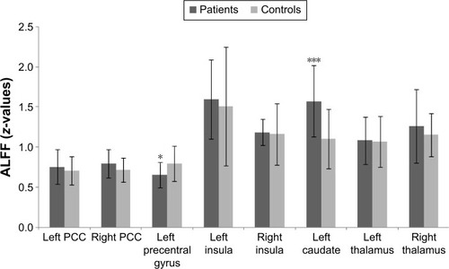
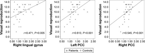
Figure 3 Correlation of regional ALFF with cognitive ability in patients with COPD and controls.
Abbreviations: ALFF, amplitude of low-frequency fluctuation; COPD, chronic obstructive pulmonary disease; PCC, posterior cingulate cortex.