Figures & data
Figure 1 Rbm and LP vessels stained with anti-Collagen IV antibody in (A) and anti-Factor VIII antibody in (B). The epithelium sits on the basement membrane. The thickness of the Rbm, is shown with the two-headed arrow. Vessels are in contact or embedded within the Rbm (arrows). Arrowheads indicate vessels in the lamina propria. Magnification ×400; scale bar =20 µm.
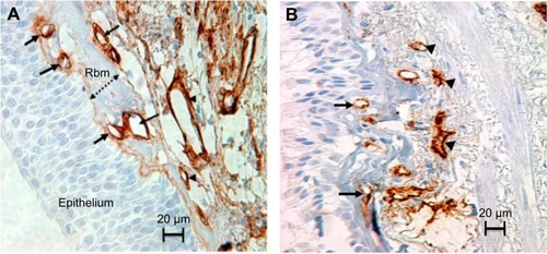
Figure 2 Study subjects and design.
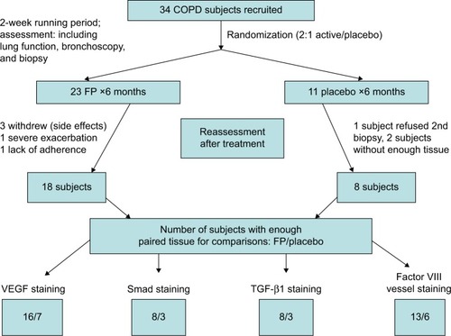
Table 1 Demographics of the study groupsTable Footnotea
Table 2 Comparison of baseline tissue vascular parameters between groupsTable Footnotea
Figure 3 The effects of ICS or placebo on LP vessels. The box plot of H-N shows that the LP was hypovascular in both treatment groups. Bars indicate medians. Dots and triangles represent current smoking and ex-smoking COPD subjects. There was strong trend for an increase in vessels overall, but this was confined to active smokers in whom the change was significant (P=0.05).
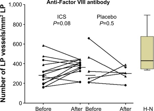
Table 3 Changes with treatmentTable Footnotea
Figure 4 Significant correlation between the baseline number and change in LP vessels with ICS (fluticasone propionate) (r=−0.7, P=0.01). Circles and triangles present current smoking and ex-smoking COPD subjects, respectively.
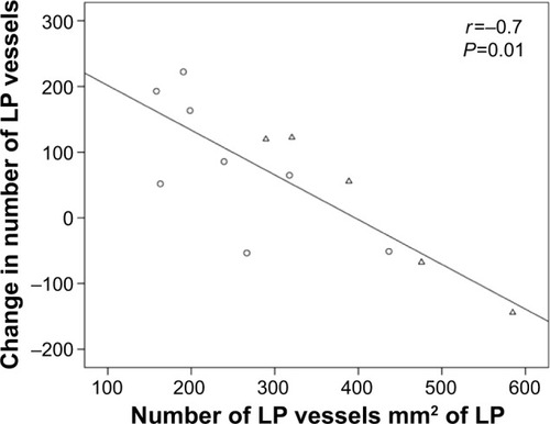
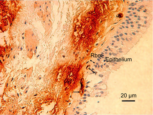
Figure S1 VEGF-stained vessels in the Rbm and LP are pointed out with narrow arrows and wide arrows, respectively.
Notes: The lamina propria is situated beneath the Rbm. The width of the Rbm is shown by a two-headed arrow. Magnification ×400.
Abbreviations: Rbm, reticular basement membrane; LP, lamina propria.
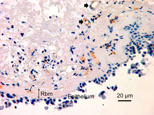
Figure S2 TGF-β-stained vessels in the Rbm are pointed out with arrows. The generalized dark immunostaining in the lamina propria, which is situated beneath the Rbm, impedes vessel identification. The width of the Rbm is shown by a two-headed arrow. Magnification ×400.
Abbreviation: Rbm, reticular basement membrane.