Figures & data
Figure 1 Inspiratory resistive breathing (IRB) at a load of 50% of maximum induces acute lung injury.
Notes: (A) Following 3 and 6 hours of IRB, increased clearance of intratracheally administered 99m Tc-DTPA from the lung is noticed, as a result of increased epithelial permeability (representative figures of 99m Tc-DTPA scintigraphy over time: upper, control; middle, 3 hours IRB; lower, 6 hours IRB; red denotes strong signal of the radiotracer). (B) IRB deranges respiratory system mechanics by decreasing static compliance after both 3 and 6 hours, as assessed by static pressure–volume (P–V) curves. Note the downward shift of the P–V curve after both 3 and 6 hours of IRB, as seen in the presence of lung injury. (C) IRB is associated with decreased PaO2:FiO2 ratio, both after 3 and 6 hours, indicating the presence of impaired gas exchange. (D) Following 6 hours of IRB, increased bronchoalveolar lavage (BAL) cellularity is noticed, due to increased counts of both macrophages and neutrophils (and lymphocytes to a lesser degree). (E) The induction of pulmonary inflammation after 6 hours of IRB is also confirmed by the presence of increased protein levels of various inflammatory cytokines in the lung tissue. (F) Representative figures of H&E-stained lung sections showing the presence of acute lung injury after 6 hours of IRB (right), compared to control (left). Note the high degree of membrane thickening, intraalveolar hemorrhage, capillary congestion, and inflammatory cell infiltration in animals that underwent 6 hours of IRB. (G) Upper: gelatin zymography of lung tissue samples revealed increased levels of MMP-9 following 6 hours of IRB, compared to control (gelatinolytic activity is visualized as white bar against blue background). Lower: alveolar macrophages and the pulmonary epithelium were the main sources of MMP-9 production after 6 hours of IRB, as detected by immunohistochemistry (representative figures of lung tissue sections, where brown denotes the presence of MMP-9). *P<0.05 to ctr; #P<0.05 to 3 hours IRB. (A)–(F) Reprinted with permission of the American Thoracic Society. Copyright © 2016 American Thoracic Society. Toumpanakis D, Kastis GA, Zacharatos P, et al.Citation9 Inspiratory resistive breathing induces acute lung injury. Am J Respir Crit Care Med. 2010;182:1129–1136. The American Journal of Respiratory and Critical Care Medicine is an official journal of the American Thoracic Society. (G) Reproduced with permission from Toumpanakis D, Noussia O, Sigala I, et al. Inspiratory resistive breathing induces MMP-9 and MMP-12 expression in the lung. Am J Physiol Lung Cell Mol Physiol. 2015;308:L683–L692.Citation10
Abbreviations: DTPA, diethylene-triamine-pentaacetate; ctr, control; hrs, hours; MIP, macrophage inflammatory protein; TNF-α, tumor necrosis factor alpha; IL, interleukin; H&E, hematoxylin and eosin; MMP, matrix metalloproteinase.
Abbreviations: DTPA, diethylene-triamine-pentaacetate; ctr, control; hrs, hours; MIP, macrophage inflammatory protein; TNF-α, tumor necrosis factor alpha; IL, interleukin; H&E, hematoxylin and eosin; MMP, matrix metalloproteinase.
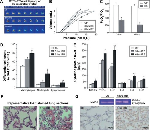
Figure 2 Resistive breathing (RB) in mice and the role of soluble guanylate cyclase.
Notes: RB in mice: (A) Tracheal banding induces lung injury. Lung histological evaluation by light microscopy revealed the existence of neutrophil infiltration in mice 24 hours after tracheal banding. RB increases interstitial and intraalveolar infiltration and focal congestion in the lung tissue. Representative histological section stained with hematoxylin and eosin of quietly breathing (control) and treated mice, after tracheal banding, respectively (upper panel). (B) Tracheal banding induces inflammatory cells influx in the BALF. Total cells and macrophages in BALF of TB mice are increased compared to sham-operated mice (control) following 24 hours of resistive breathing. Respiratory system mechanics: (C) Airway resistance by force oscillation technique is elevated following tracheal banding. (D) The expression of sGC is decreased in the lungs of TB mice. Protein levels of sGC subunits are decreased in mice after 24 hours of resistive breathing. Sham-operated (control) or TB mice were sacrificed 24 hours after treatment. Representative Western blots for α-1, β-1, and β-actin are presented (upper panel). Blots were quantified by densitometry. Expression for each subunit normalized for β-actin was set at 100% for sham-operated mice. (E) Therapeutic activation of sGC attenuates lung injury caused by tracheal banding. Lung histological evaluation by light microscopy revealed the attenuation of inflammation and neutrophil infiltration in mice treated with BAY58-2667. BAY58-2667 decreased interstitial and intraalveolar infiltration and focal congestion in the lung tissue, compared to the group of TB mice. (F) Activation of sGC improves lung mechanics after tracheal banding. Elevated airway resistance, following tracheal banding, was attenuated when BAY58-2667 was administrated after TB. Values are expressed as mean ± SEM; n=10; *P<0.05 for sham-operated (control) group and #P<0.05 for TB + BAY58-2667 group. Figures A–F have been reprinted with permission of the American Thoracic Society. Copyright © 2016 American Thoracic Society. Glynos C, Toumpanakis D, Loverdos K, et al. Guanylyl cyclase activa tion reverses resistive breathing-induced lung injury and inflammation. Am J Respir Cell Mol Biol. 2015;52:762–771.Citation23 The American Journal of Respiratory Cell and Molecular Biology is an official journal of the American Thoracic Society. Smoking mice: (G) sGC expression is decreased in mice after cigarette smoke (CS) exposure. Protein levels of sGCα-1 upon acute (3 days), subacute (4 weeks), and chronic (24 weeks) CS exposure. Example of Western blot for sGCα-1 is presented (upper panel). Western blot was performed on eight mice per group. Blots were quantified by densitometry. Expression for the sGCα-1 subunit normalized for β-actin was set at 100% for air-exposed mice. Values are expressed as mean ± SEM; n=8/group; *P<0.05, **P<0.01 for air-exposed mice. (H) BAY58-2667 administration attenuates airway hyperresponsiveness on acute CS exposure. BAY58-2667 administration in acute CS-exposed mice ameliorates bronchoconstriction. Effect of BAY58-2667 on percent increase in airway resistance (R) in a dose–response manner to serotonin (5-HT) challenge is presented. Values are expressed as mean ± SEM; n=8/group; *P<0.05 for air-exposed group and #P<0.05 for CS + BAY58-2667 group. COPD patients and smokers: (I) sGC protein expression is decreased in patients with COPD. Western blot was performed on a total of 33 patients (nine never smokers, nine smokers, and 15 patients with COPD). Examples of Western blots for sGCα-1 and β-actin are presented (upper panel). Expression for the sGCα-1 subunit normalized for β-actin was set at 100% for NS. Values are expressed as mean ± SEM; *P<0.05. (J) mRNA expression of the sGCα-1 subunit correlates with lung function in smokers and patients with COPD. sGC mRNA levels of α-1 subunit of human lung tissues were correlated with forced expiratory volume in 1 second (FEV1%) (% of predicted). Spearman correlation coefficient (rs) and P-value are shown. Figures G–J have been reprinted with permission of the American Thoracic Society. Copyright © 2016 American Thoracic Society. Glynos C, Dupont LL, Vassilakopoulos T, et al. The role of soluble guanylyl cyclase in chronic obstructive pulmonary disease. Am J Respir Crit Care Med. 2013;188:789–799.Citation25 The American Journal of Respiratory and Critical Care Medicine is an Official Journal of the American Thoracic Society.
Abbreviations: TB, tracheal banding; BALF, bronchoalveolar lavage fluid; sGC, soluble guanylyl cyclase; SEM, standard error of the mean; NS, never smokers; S, smokers; wks, weeks.
Abbreviations: TB, tracheal banding; BALF, bronchoalveolar lavage fluid; sGC, soluble guanylyl cyclase; SEM, standard error of the mean; NS, never smokers; S, smokers; wks, weeks.
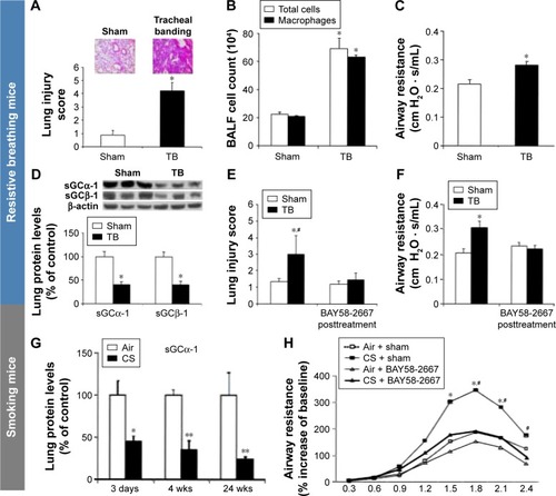
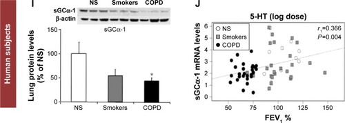
Figure 3 Inspiratory resistive breathing (IRB) induces IL-6 upregulation in the induced sputum of healthy humans.
Notes: Five healthy nonsmoking volunteers gave an induced sputum sample on the morning of day 1. Three days later in the morning, they were under inspiratory resistive breathing at 50% of maximum inspiratory pressure for 45 minutes. After that resistive breathing-induced sputum was collected. Interleukin 6 (IL-6) was determined in the sputum supernatant by ELISA. Data are presented as mean ± SEM; *P=0.038 before RB.
Abbreviations: ELISA, enzyme-linked immunosorbent assay; SEM, standard error of the mean; RB, resistive breathing.
Abbreviations: ELISA, enzyme-linked immunosorbent assay; SEM, standard error of the mean; RB, resistive breathing.
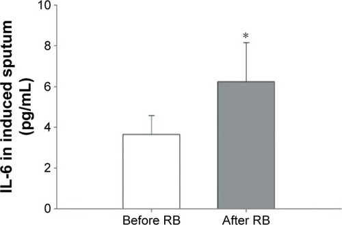
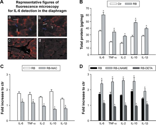
Figure 4 Inspiratory resistive breathing (IRB) induces cytokine upregulation in the diaphragm: the role of oxidative stress and nitric oxide (NO).
Notes: (A) Localization of interleukin 6 (IL-6) protein expression in rat diaphragms. Antibodies showed positive IL-6 staining in diaphragms of rats subjected to 6 hours of IRB. Membrane-associated (white arrows in a) and punctuate cytosolic (white arrows in b) positive IL-6 staining were evident inside small muscle fibers, whereas large muscle fibers showed no IL-6 staining (blue arrows). Blood vessels were negative for IL-6 protein (white arrow in c). Very weak IL-6 staining was detectable in the diaphragm of quietly breathing rats (d). Reprinted with permission of the American Thoracic Society. Copyright © 2016 American Thoracic Society. Vassilakopoulos T, Divangahi M, Rallis G, et al. Differential cytokine gene expression in the diaphragm in response to strenuous resistive breathing. Am J Respir Crit Care Med. 2004;170:154–161.Citation8 The American Journal of Respiratory and Critical Care Medicine is an Official Journal of the American Thoracic Society. (B) IRB induces cytokine expression in the diaphragm of rats. Cumulative results of ELISA depicting the protein levels of IL-6, TNF-α, IL-2, IL-10, and IL-1β in the diaphragm of quietly breathing rats (Ctr) and rats subjected to IRB for 6 hours (RB). Data are presented as mean ± SE pg/mg total protein (*P<0.05 vs control). Modified from Sigala et al.Citation30 (C), (D) Effect of antioxidant (NAC) administration (C) and NO production modification with the nonselective nitric oxide synthase (NOS) inhibitor (L-NAME) and the NO donor (DETA- NONOate) (D) on IRB-induced cytokine upregulation in the diaphragm. Protein levels (estimated with ELISA) of IL-6, tumor necrosis factor alpha (TNF-α), IL-2, IL-10, and IL-1β, respectively, in the diaphragm of rats subjected to IRB for 6 hours treated with NAC (RB-NAC) or not (RB) (C) and with NOS inhibitor (RB-LNAME), NO donor (RB-DETA), or nothing (RB) (D). Data are presented as fold increase to control (mean ± SE; *P<0.05 vs RB). Reproduced with permission from Sigala I, Zacharatos P, Toumpanakis D, et al. MAPKs and NF-kappaB differentially regulate cytokine expression in the diaphragm in response to resistive breathing: the role of oxidative stress. Am J Physiol Regul Integr Comp Physiol. 2011;300:R1152–R1162;Citation30 and Sigala I, Zacharatos P, Boulia S, et al. Nitric oxide regulates cytokine induction in the diaphragm in response to inspiratory resistive breathing. J Appl Physiol (1985). 2012;113:1594–1603.Citation31
Abbreviations: ELISA, enzyme-linked immunosorbent assay; Ctr, control; SE, standard error; RB, resistive breathing; NAC, N-acetylcysteine.
Abbreviations: ELISA, enzyme-linked immunosorbent assay; Ctr, control; SE, standard error; RB, resistive breathing; NAC, N-acetylcysteine.