Figures & data
Figure 1 CSE increases PlGF expression and secretion in 16-HBE cells.
Notes: Cells were treated with or without CSE at the designed concentrations and time intervals. Media samples and cells were collected and the mRNA, protein and secretion of PlGF was assessed by qPCR (A and B), Western blot (C and D) and ELISA (E and F). Cell viability was assessed by CCK-8 assay (G). All experiments were repeated in triplicate. *P<0.05.
Abbreviations: CSE, cigarette smoke extract; PlGF, placental growth factor; HBE, human bronchial epithelium; qPCR, quantitative polymerase chain reaction; ELISA, enzyme-linked immunosorbent assay; CCK, Cell Counting Kit-8; GAPDH, glyceraldehyde 3-phosphate dehydrogenase; CTL, control.
Abbreviations: CSE, cigarette smoke extract; PlGF, placental growth factor; HBE, human bronchial epithelium; qPCR, quantitative polymerase chain reaction; ELISA, enzyme-linked immunosorbent assay; CCK, Cell Counting Kit-8; GAPDH, glyceraldehyde 3-phosphate dehydrogenase; CTL, control.
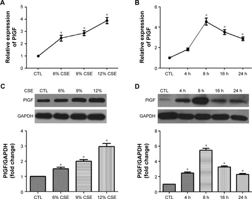
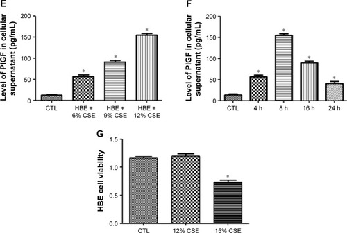
Figure 2 CSE increases of Egr-1 expression, nuclear accumulation and transcriptional activity in 16-HBE cells.
Notes: Cells were treated with or without CSE at the designed concentrations (3%, 6%, 9%, 12%) and time intervals (1 h, 2 h, 4 h, 8 h, 16 h). Total RNA or whole cell lysates were collected, and the expression of Egr-1 was assessed by qPCR (A and B) and Western blot (C and D). Nuclear localization of Egr-1 was examined by immunofluorescence staining (E). Dose–response of the transcriptional activity of Egr-1 was assayed with nuclear extracts using the BD Mercury TransFactor kit (F). All experiments were repeated in triplicate. *P<0.05.
Abbreviations: CSE, cigarette smoke extract; Egr-1, early growth response-1; HBE, human bronchial epithelium; qPCR, quantitative polymerase chain reaction; GAPDH, glyceraldehyde 3-phosphate dehydrogenase; CTL, control.
Abbreviations: CSE, cigarette smoke extract; Egr-1, early growth response-1; HBE, human bronchial epithelium; qPCR, quantitative polymerase chain reaction; GAPDH, glyceraldehyde 3-phosphate dehydrogenase; CTL, control.
Figure 3 siRNA for Egr-1 suppresses CSE-induced Egr-1 and PlGF mRNA and protein expression.
Notes: After transfection with 150 nM Egr-1 siRNA-1, siRNA-2 or scrambled siRNA for 24 h, the cells were exposed to CSE (12%); 16-HBE cells transfected with Egr-1 siRNA showed a significant downregulation in Egr-1 mRNA (A), Egr-1 protein (B), PlGF mRNA (C) and PlGF protein (D). All experiments were repeated in triplicate. *P<0.05.
Abbreviations: Egr-1, early growth response-1; CSE, cigarette smoke extract; PlGF, placental growth factor; HBE, human bronchial epithelium; GAPDH, glyceraldehyde 3-phosphate dehydrogenase; CTL, control.
Abbreviations: Egr-1, early growth response-1; CSE, cigarette smoke extract; PlGF, placental growth factor; HBE, human bronchial epithelium; GAPDH, glyceraldehyde 3-phosphate dehydrogenase; CTL, control.
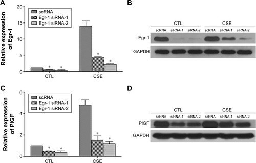
Figure 4 CSE increases the phosphorylation and nuclear translocation of the ERK-1/2 in 16-HBE cells.
Notes: Cells were exposed to CSE (0–240 min), and cell lysates was subjected to Western blot analysis for ERK-1/2 phosphorylation measurement (A and B). Nuclear accumulation of phospho-ERK-1/2 in CSE-treated cells (0–60 min) was detected by immunofluorescence staining (C). Cells were pretreated with U0126 (10 μmol/L, 1 h) and then CSE was added (4 h). Egr-1 and PlGF levels were examined by Western blot analysis (D). All experiments were repeated in triplicate. *P<0.05 compared with normal control; #P<0.05 compared with CSE treated control.
Abbreviations: CSE, cigarette smoke extract; ERK-1/2, extracellular signal-regulated kinase1/2; HBE, human bronchial epithelium; Egr-1, early growth response-1; PlGF, placental growth factor; GAPDH, glyceraldehyde 3-phosphate dehydrogenase; CTL, control.
Abbreviations: CSE, cigarette smoke extract; ERK-1/2, extracellular signal-regulated kinase1/2; HBE, human bronchial epithelium; Egr-1, early growth response-1; PlGF, placental growth factor; GAPDH, glyceraldehyde 3-phosphate dehydrogenase; CTL, control.
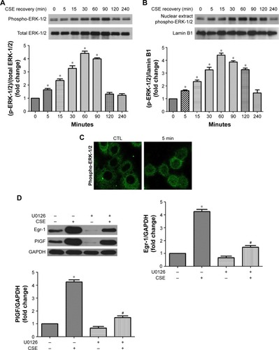
Figure 5 Effects of CSE on the ROS surge in 16-HBE cells.
Notes: Cells were pretreated with or without NAC (10 mmol/L) for 2 h, followed by addition of CSE (60 min). Subsequently, cellular ROS levels were measured by CM-H2DCFDA assay (A), the activity of SOD was detected by SOD assay (B), phospho-ERK-1/2 was measured by Western blot analysis (C) and Egr-1 and PlGF expression were measured by qRT-PCR (D). All experiments were repeated in triplicate. *P<0.05 compared with control. #P<0.05 compared with CSE treated.
Abbreviations: CSE, cigarette smoke extract; ROS, reactive oxygen species; HBE, human bronchial epithelium; NAC, N-acetyl-L-cysteine; CM-H2DCFDA, 5-(and-6)-chloromethyl-2′,7′-dichlorodihydrofluorescein diacetate; SOD, superoxide dismutase; ERK-1/2, extracellular signal-regulated kinase-1/2; Egr-1, early growth response-1; PlGF, placental growth factor; qRT-PCR, quantitative reverse transcription polymerase chain reaction; DCF, 2′,7′-dichlorodihydrofluorescein; CTL, control.
Abbreviations: CSE, cigarette smoke extract; ROS, reactive oxygen species; HBE, human bronchial epithelium; NAC, N-acetyl-L-cysteine; CM-H2DCFDA, 5-(and-6)-chloromethyl-2′,7′-dichlorodihydrofluorescein diacetate; SOD, superoxide dismutase; ERK-1/2, extracellular signal-regulated kinase-1/2; Egr-1, early growth response-1; PlGF, placental growth factor; qRT-PCR, quantitative reverse transcription polymerase chain reaction; DCF, 2′,7′-dichlorodihydrofluorescein; CTL, control.
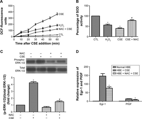
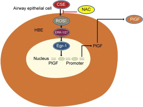
Figure 6 A schematic representation depicting the ROS/ERK/Egr-1 axis involved in CSE-induced PlGF expression in airway epithelial cells.
Abbreviations: ROS, reactive oxygen species; ERK, extracellular signal-regulated kinase; Egr-1, early growth response-1; CSE, cigarette smoke extract; PlGF, placental growth factor; HBE, human bronchial epithelium; NAC, N-acetyl-L-cysteine.