Figures & data
Table 1 Demographic and baseline details of subjects, including symptoms and functional characteristics
Table 2 Association between bronchiectasis, gender, severe COPD symptoms (CAT ≥10), more dyspnea, severity of airflow limitation and frequent COPD exacerbation (≥2 in the past year) and/or hospitalized exacerbation in the past year
Table 3 Symptoms and physiological conditions of COPD patients with and without bronchiectasis
Table 4 Proportion of bacterial and mycobacterial pathogens isolated from the sputum of COPD patients, including isolated pathogens in patients with and without frequent or severe exacerbation
Table 5 Other CT findings among COPD patients
Figure 1 Axial CT image with a lung-window setting showing diffuse emphysema with varicose bronchiectasis in the right lower lobe and tubular bronchiectasis in the left lower lobe.
Abbreviation: CT, computed tomography.
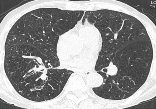
Figure 2 Axial CT images with a lung-window setting showing tubular bronchiectasis with the signet ring sign in the right lower lobe (A–C) and the tram-track sign in the left lower lobe (B).
Abbreviation: CT, computed tomography.
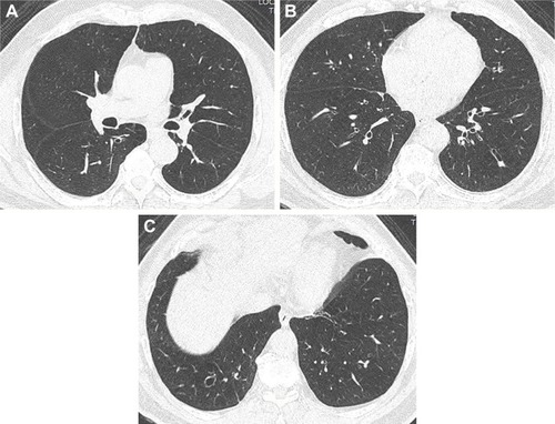
Figure 3 Axial CT images with a lung-window setting showing combined tubular and varicose bronchiectasis, emphysema and fibrosis in bilateral upper lobes (A), right middle lobe (B) and right lower lobe (B).
Abbreviation: CT, computed tomography.
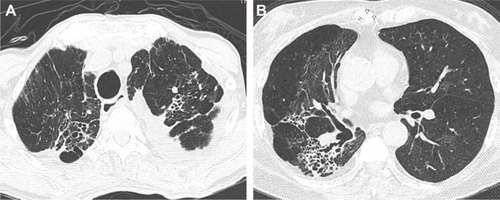
Figure 4 Axial CT images with a lung-window setting showing severe centrilobular emphysema and bronchiectasis in both lungs (A–C).
Abbreviation: CT, computed tomography.
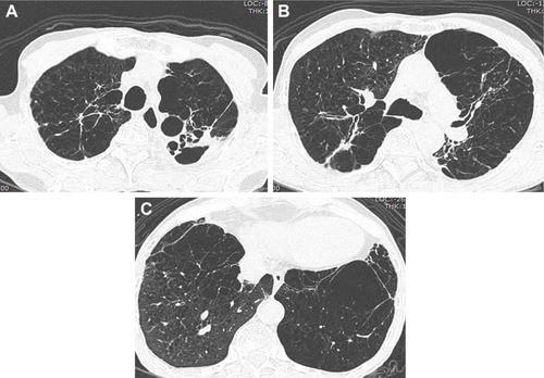
Figure 5 Axial CT image with a lung-window setting showing emphysema and bronchial wall thickening in bilateral lower lobes.
Abbreviation: CT, computed tomography.
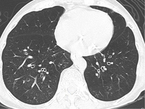
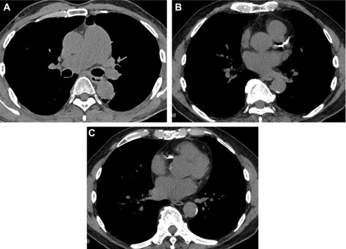
Figure 6 Axial CT images with a mediastinal-window setting showing a dilated main pulmonary artery (A) and calcifications of the aorta and coronary arteries (B–C).