Figures & data
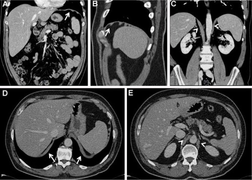
Figure 1 CT images.
Notes: The CT images in the coronal and axial planes allow visualization of the diaphragm as a hyperdense linear band interposed between the chest and the abdominal cavity (A and D, respectively; see arrows). Sagittal images highlight a sort of “corrugated” morphology that shows the orientation of the muscle bundles (B; see arrow), which may appear more or less pronounced in wellness or pathologic conditions, such as COPD. Clearly visible diaphragmatic pillars also appear in both the coronal plane (C) and the axial plane (E) (arrowheads).
Abbreviation: CT, computed tomography.
Abbreviation: CT, computed tomography.