Figures & data
Table 1 Clinical characteristics and spirometric values
Figure 1 A flowchart for patient selection.
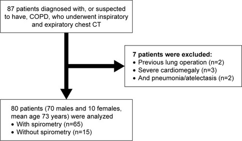
Figure 2 An example of tracheobronchial angle measurements by the research software.
Abbreviations: LMB, left main bronchus; RMB, right main bronchus.
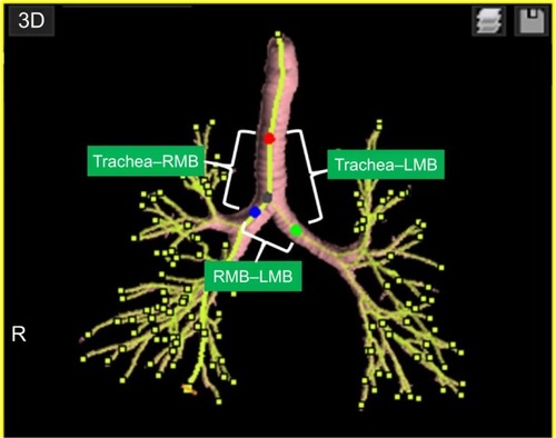
Table 2 Tracheobronchial angles and LV measurements
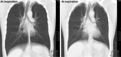
Figure 3 Reconstructed coronal CT images of a 72-year-old male smoker with mild COPD (FEV1/FVC=66%, LAV%=13%).
Abbreviations: COPD, chronic obstructive pulmonary disease; CT, computed tomography; FEV1, forced expiratory volume in 1 second; FVC, forced vital capacity; LAV, low attenuation volume; LMB, left main bronchus; RMB, right main bronchus.
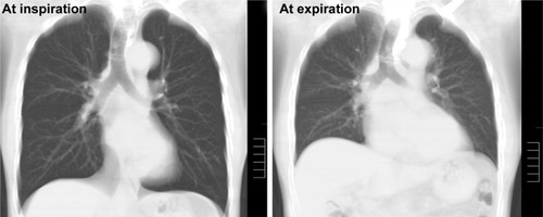
Table 3 Correlations between tracheobronchial angles and LV measurements
Table 4 Correlations between tracheobronchial angles and COPD indices
Figure 4 Correlations between tracheobronchial angles and spirometric values.
Notes: The angles formed by the right and left main bronchi (RMB–LMB) at inspiration and expiration significantly correlate with spirometric values (FEV1/FVC).
Abbreviations: FEV1, forced expiratory volume in 1 second; FVC, forced vital capacity; LMB, left main bronchus; RMB, right main bronchus.
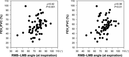
Figure 5 Reconstructed coronal CT images of a 54-year-old male with severe COPD (FEV1/FVC=42%, LAV%=43%).
Abbreviations: COPD, chronic obstructive pulmonary disease; CT, computed tomography; FEV1, forced expiratory volume in 1 second; FVC, forced vital capacity; LAV, low attenuation volume; LMB, left main bronchus; RMB, right main bronchus.