Figures & data
Table 1 Anthropometric data and pulmonary function (patients and controls)
Table 2 Sleep quality and sleep disordered breathing at baseline
Table 3 Treatment effects of NHF and supplemental O2 on sleep, oxygenation, and sleep disordered breathing
Table 4 AA indices during REM sleep, NREM sleep, and the NREM sleep stages N1, N2, and N3
Figure 1 Example of changes in PWA during REM sleep in one COPD patient on three different conditions: RA, O2 supplementation, and NHF treatment.
Notes: Autonomic activation events were defined as a reduction in PWA of 30% or more compared to a prior baseline period. Automatically scored autonomic activation events are highlighted in the pulse wave tracings.
Abbreviations: AAI, autonomic activation index; EEG, electroencephalogram; EMG, electromyogram; EOG, electrooculogram; NHF, nasal high flow; PWA, pulse wave amplitude; RA, room air; REM, rapid eye movement.
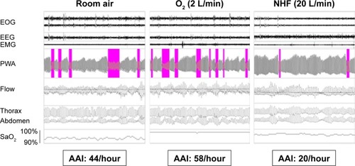
Figure 2 Modification of vascular AAI with nasal high flow and supplemental O2 compared to the room air condition during REM sleep.
Abbreviations: AAI, autonomic activation index; RA, room air; REM, rapid eye movement.
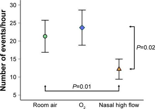
Figure 3 Correlation between the reduction of vascular AAI during REM sleep by NHF compared with the room air condition (y-axis) and impaired lung function assessed as FEV1 (x-axis).
Note: Note that a reduction of REM-AAI during NHF treatment translates to a positive value on the y-axis and vice versa.
Abbreviations: AAI, autonomic activation index; NHF, nasal high flow; REM, rapid eye movement.
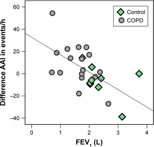
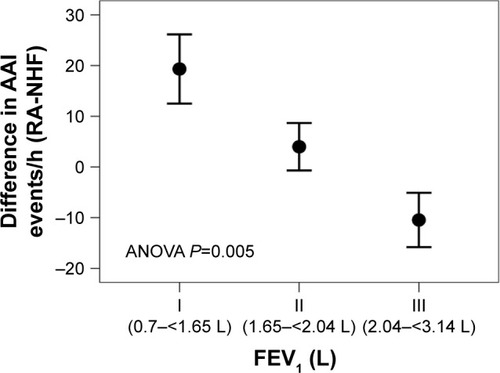
Figure 4 Relation between FEV1 (tertiles I–III) and the difference in sympathetic activity (AAI) between the room air condition and NHF during REM sleep.
Abbreviations: AAI, autonomic activation index; NHF, nasal high flow; REM, rapid eye movement.