Figures & data
Table 1 Demographics on the Volunteers
Figure 1 RELMβ expression was significantly increased in patients with COPD. (A) RELMβ mRNA expression in patients with COPD and normal controls in the GEO database. (B) RELMβ protein expression in the sera of patients with COPD and control individuals.*p≤0.05, **p≤0.01, ****p≤0.0001.
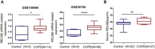
Figure 2 RELMβ regulates inflammatory networks. (A) The online data analysis site STRING was used to analyze the inflammatory networks involved in the regulation of RELMβ expression. (B-C) Results of RELMβ and provocative factor enrichment analysis. (D-G) Correlation of high and low expression of RELMβ mRNA with the expression of the inflammatory factors IL-8 and IL-1β. *p≤0.05.
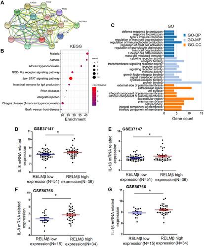
Figure 3 RELMβ promotes the expression of the inflammatory factors IL-8 and IL-1β. (A1, A2) 16HBE cells were stimulated with different concentrations of recombinant RELMβ protein; Western blotting results showed the expression of the inflammatory factors IL-8 and IL-1β. (B1, B2) ELISA results showed the secretion of the inflammatory factors IL-8 and IL-1β into cell supernatants. (C1–C3) Using RT-PCR and Western blotting, we found that RELMβ expression was increased in 16HBE cells infected with lentiviral particles compared with parental cells and cells infected with empty vector at both the mRNA and protein levels. (D1–D3) We found that in the RELMβ-overexpressing cell strain, the inflammatory factors IL-8 and IL-1β were expressed at higher levels than those in the parental and null control cell strains at both the mRNA and protein levels. (E1–E3) We successfully knocked down RELMβ gene expression; even under stimulation with tobacco smoke extract, the expression of RELMβ at both the mRNA and protein levels was lower than that in the tobacco smoke extract treatment group. (F1, F2) We found that the expression of the inflammatory factors IL-8 and IL-1β was significantly downregulated after successful knock down of RELMβ gene expression. *p≤0.05, **p≤0.01, ***p≤0.001, ****p≤0.0001.
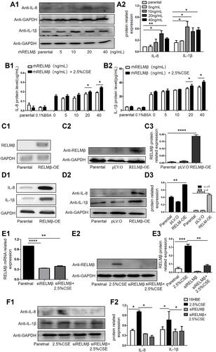
Figure 4 RELMβ activates the phosphorylated p38 MAPK signaling pathway. (A-C) Western blotting results showed that stable overexpression of RELMβ in 16HBE cells could not induce the phosphorylation of ERK1/2, Akt, and SAPK/JNK. (D1, D2) Our Western blotting results showed that RELMβ overexpression could induce phosphorylated p38 MAPK expression. (E1, E2) When we knocked down RELMβ gene expression, phosphorylated p38 MAPK was still not activated even after stimulation with tobacco smoke extract. *p≤0.05.
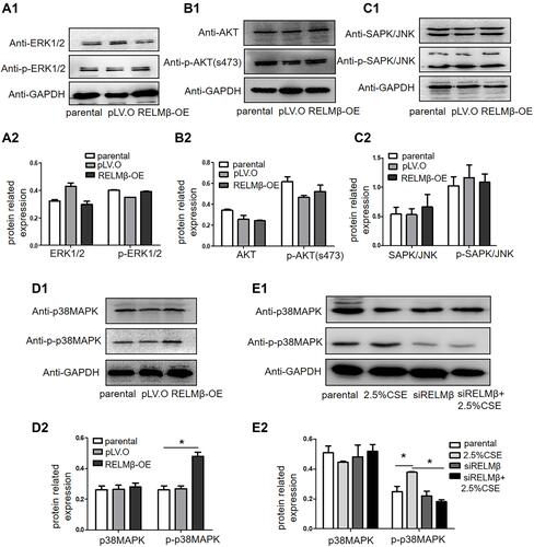
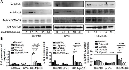
Figure 5 RELMβ promotes the expression of the inflammatory factors IL-8 and IL-1β via the p38 MAPK signaling pathway. (A-B) Western blotting and bar graphs showing the expression of p-p38 MAPK, IL-8, and IL-1β in the 16HBE cells in the parental control, null and RELMβ overexpression groups after treatment with the p38 MAPK inhibitor sb203580. *p≤0.05, **p≤0.01, ***p≤0.001, ****p≤0.0001.