Figures & data
Figure 1 The chemical structure of X22.
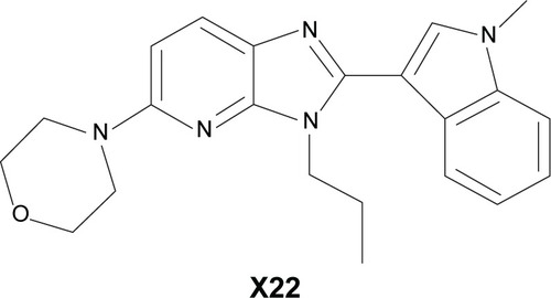
Figure 2 Effect of X22 on MPMs and RAW 264.7 macrophage viability analyzed by an MTT assay.
Notes: MPMs (50,000 cells/mL) (A) or RAW 264.7 (80,000 cells/mL) (B) macrophages were seeded in 96-well plates and treated with the indicated concentration of X22 for 24 hours. All values are given as the mean ± SEM of three independent experiments.
Abbreviations: DMSO, dimethyl sulfoxide; MPMs, mouse peritoneal macrophages; SEM, standard error of mean.
Abbreviations: DMSO, dimethyl sulfoxide; MPMs, mouse peritoneal macrophages; SEM, standard error of mean.
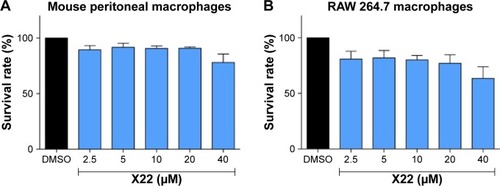
Figure 3 X22 inhibited LPS-induced inflammatory cytokine expression in vitro.
Notes: (A and B) MPMs or (C and D) RAW 264.7 macrophages were pretreated with X22 (2.5, 5, 10, 20, and 40 μM) for 30 minutes, followed by incubation with LPS (0.5 μg/mL) for 24 hours. IL-6 and TNF-α levels in the culture medium were measured by ELISA analysis and normalized to the total protein. The results are presented as the percent of LPS control. Each bar represents the mean ± SEM of three independent experiments. Statistical significance relative to the LPS group is indicated, *P<0.05; **P<0.01. (E) MPMs were pretreated with X22 (20 μM) for 30 minutes followed by incubation with LPS (0.5 μg/mL) for 6 hours. The mRNA levels of inflammatory cytokines were quantified by real-time qPCR analysis using β-actin mRNA as the internal control. Each bar represents the mean ± SEM of three to five independent experiments (*P<0.05, **P<0.01 relative to the LPS group).
Abbreviations: COX-2, cyclooxygenase-2; DMSO, dimethyl sulfoxide; ELISA, enzyme-linked immunosorbent assay; IL-6, interleukin-6; IL-1β, interleukin-1β; LPS, lipopolysaccharide; qPCR, quantitative polymerase chain reaction; TNF-α, tumor necrosis factor-α; MPMs, mouse peritoneal macrophages; SEM, standard error of mean.
Abbreviations: COX-2, cyclooxygenase-2; DMSO, dimethyl sulfoxide; ELISA, enzyme-linked immunosorbent assay; IL-6, interleukin-6; IL-1β, interleukin-1β; LPS, lipopolysaccharide; qPCR, quantitative polymerase chain reaction; TNF-α, tumor necrosis factor-α; MPMs, mouse peritoneal macrophages; SEM, standard error of mean.
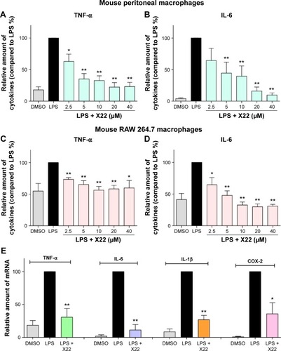
Figure 4 X22 attenuated LPS-induced septic shock in vivo.
Notes: (A) Male ICR mice were injected intravenously with vehicle or X22 (25 and 50 mg/kg) and the body weight was recorded for 7 days. (B) Male C57BL/6 mice were pretreated with X22 (tail vein injection, 20 mg/kg) or vehicle, followed by injection of LPS (tail vein injection, 20 mg/kg). Survival rates were recorded for 7 days at an interval of 24 hours after the LPS injection. n=10 animals in each group. **P<0.01 versus LPS group.
Abbreviations: CON, control; ICR mice, Institute of Cancer Research mice; LPS, lipopolysaccharide.
Abbreviations: CON, control; ICR mice, Institute of Cancer Research mice; LPS, lipopolysaccharide.
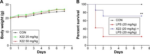
Figure 5 X22 decreased TNF-α expression in the serum of septic mice.
Notes: Male C57BL/6 mice were treated with LPS (tail vein injection, 20 mg/kg) 15 minutes before X22 (tail vein injection, 10 and 20 mg/kg) or 15 minutes after X22 (tail vein injection, 20 mg/kg) injection. Mice were anesthetized and killed 6 hours after LPS injection. The TNF-α level in serum was detected by ELISA analysis. *P<0.05, **P<0.01 versus LPS group.
Abbreviations: CON, control; ELISA, enzyme-linked immunosorbent assay; LPS, lipopolysaccharide; TNF-α, tumor necrosis factor-α.
Abbreviations: CON, control; ELISA, enzyme-linked immunosorbent assay; LPS, lipopolysaccharide; TNF-α, tumor necrosis factor-α.
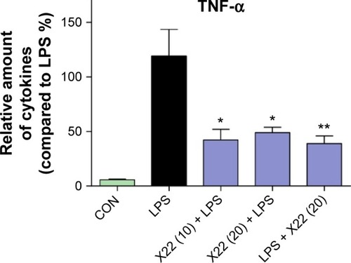
Figure 6 X22 attenuated sepsis-induced lung injury in mice.
Notes: Male C57BL/6 mice were treated with LPS (tail vein injection, 20 mg/kg) 15 minutes before X22 (tail vein injection, 10 and 20 mg/kg) or 15 minutes after X22 (tail vein injection, 20 mg/kg) injection. Mice were anesthetized and killed 6 hours after the LPS injection. The expression of inflammatory mediators in lung tissue, including TNF-α (A), IL-6 (B), iNOS (C), and IL-1β (D), was analyzed by real-time qPCR using β-actin mRNA as the internal control (*P<0.05, **P<0.01 relative to the LPS group). (E) Compound X22 attenuated sepsis-induced histopathological change in lung tissue (H&E staining, 200×). (F) The lung injury score was determined. (G) CD68 immunostaining was used for the detection of macrophage infiltration in the lung tissue, (200×).
Abbreviations: CON, control; H&E staining, hematoxylin and eosin staining; IL-6, interleukin-6; IL-1β, interleukin-1β; iNOS, inducible nitric oxide synthase; LPS, lipopolysaccharide; qPCR, quantitative polymerase chain reaction; TNF-α, tumor necrosis factor-α.
Abbreviations: CON, control; H&E staining, hematoxylin and eosin staining; IL-6, interleukin-6; IL-1β, interleukin-1β; iNOS, inducible nitric oxide synthase; LPS, lipopolysaccharide; qPCR, quantitative polymerase chain reaction; TNF-α, tumor necrosis factor-α.
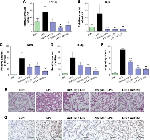
Figure 7 X22 attenuated sepsis-induced liver injury in mice.
Notes: Male C57BL/6 mice were treated with LPS (tail vein injection, 20 mg/kg) 15 minutes before X22 (tail vein injection, 10 and 20 mg/kg) or 15 minutes after X22 (tail vein injection, 20 mg/kg) injection. Mice were anesthetized and killed 6 hours after the LPS injection. (A–D) The expression of the inflammatory mediators in liver tissue, including TNF-α (A), IL-6 (B), iNOS (C), and IL-1β (D), was analyzed by real-time qPCR using β-actin mRNA as the internal control (*P<0.05, **P<0.01). X22 lowered serum ALT (E) and serum AST (F) expression in septic mice (*P<0.05, **P<0.01 relative to the LPS group). (G) Compound X22 attenuated the sepsis-induced histopathological change in liver tissue (H&E staining, 200×). (H) The liver injury score was determined based on “G”. (I) CD68 immunostaining was used for the detection of macrophage infiltration in the liver tissue, (200×).
Abbreviations: ALT, alanine aminotransferase; AST, aspartate transaminase; CON, control; H&E staining, hematoxylin and eosin staining; IL-6, interleukin-6; IL-1β, interleukin-1β; LPS, lipopolysaccharide; iNOS, inducible nitric oxide synthase; qPCR, quantitative polymerase chain reaction; TNF-α, tumor necrosis factor-α.
Abbreviations: ALT, alanine aminotransferase; AST, aspartate transaminase; CON, control; H&E staining, hematoxylin and eosin staining; IL-6, interleukin-6; IL-1β, interleukin-1β; LPS, lipopolysaccharide; iNOS, inducible nitric oxide synthase; qPCR, quantitative polymerase chain reaction; TNF-α, tumor necrosis factor-α.
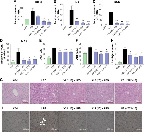
Figure 8 Effect of X22 on LPS-induced acute lung injury.
Notes: Mice were treated by intratracheal instillation of LPS. Six hours later, mice were anesthetized and killed. Lung tissues and BALF were collected for further tests. (A) Compound X22 attenuated LPS-induced histopathological change in lung tissue (H&E staining, 200×) (magnification: ×200, scale bar: 100 μm). (B) The lung injury score was determined. (C–F) Effects of X22 on total cells in BALF (C), TNF-α (D), and IL-6 (E) levels in BALF, and TNF-α (F) levels in serum (*P<0.05, **P<0.01 relative to the LPS group). (G–I) The expression of inflammatory mediators in lung tissue, including IL-6 (G), IL-1β (H), and COX-2 (I), was analyzed by real-time qPCR using β-actin mRNA as the internal control (*P<0.05, **P<0.01 relative to the LPS group).
Abbreviations: BALF, bronchoalveolar lavage fluid; CON, control; COX-2, cyclooxygenase-2; H&E staining, hematoxylin and eosin staining; IL-6, interleukin-6; IL-1β, interleukin-1β; LPS, lipopolysaccharide; qPCR, quantitative polymerase chain reaction; TNF-α, tumor necrosis factor-α.
Abbreviations: BALF, bronchoalveolar lavage fluid; CON, control; COX-2, cyclooxygenase-2; H&E staining, hematoxylin and eosin staining; IL-6, interleukin-6; IL-1β, interleukin-1β; LPS, lipopolysaccharide; qPCR, quantitative polymerase chain reaction; TNF-α, tumor necrosis factor-α.
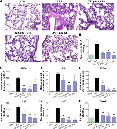
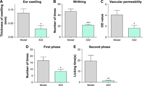
Figure 9 Effects of X22 on chemically-induced inflammation in vivo.
Notes: (A) Ear swelling induced by dimethyl benzene. (B) Acetic acid-induced writhing response. (C) Acetic acid-induced vascular permeability. Formalin-induced nociception mice, (D) first-inflammatory phase, 0–10 minutes after formalin induction, and (E) second-inflammatory phase, 15–30 minutes after formalin induction. Control animals received saline (0.9% NaCl). Each value represents the mean ± SEM of six mice, and asterisks indicate the significant difference of pretreated group and model group to the corresponding saline group (*P<0.05, **P<0.01, ***P<0.01 relative to the LPS group).
Abbreviations: LPS, lipopolysaccharide; OD value, optical density value; SEM, standard error of mean.
Abbreviations: LPS, lipopolysaccharide; OD value, optical density value; SEM, standard error of mean.