Figures & data
Figure 1 FGF1V–vcMMAE conjugate.
Note: Scheme of the FGF1V–vcMMAE conjugate with a valine–citrulline linker designed to be specifically cleaved by lysomal protease cathepsin B.
Abbreviations: FGF1V, fibroblast growth factor 1 variant; FGFR, fibroblast growth factor receptor; MMAE, monomethyl auristatin E; vcMMAE, valine–citrulline monomethyl auristatin E.
Abbreviations: FGF1V, fibroblast growth factor 1 variant; FGFR, fibroblast growth factor receptor; MMAE, monomethyl auristatin E; vcMMAE, valine–citrulline monomethyl auristatin E.

Figure 2 FGF1V–vcMMAE conjugation.
Notes: (A–C) Electrophoretic separation (left), MALDI-MS spectra (center), and FGF1 tryptophan fluorescence spectra (right) of conjugation reactions performed in different conditions and yielding a mixture of unmodified, singly, doubly, and triply labeled FGF1V in different proportions. (D) Endogenous Cys117 of FGF1 is not efficiently modified with vcMMAE at 4°C. FGF1C117 is a variant used with the stabilizing mutations and N-terminal linker (Cys117 was not mutated).
Abbreviations: CR, conjugation reaction sample; FGF1V, fibroblast growth factor 1 variant; M, molecular weight marker; MALDI-MS, matrix-assisted laser desorption-mass spectrometry; SDS-PAGE, sodium dodecyl sulfate polyacrylamide gel electrophoresis; vcMMAE, valine–citrulline monomethyl auristatin E.
Abbreviations: CR, conjugation reaction sample; FGF1V, fibroblast growth factor 1 variant; M, molecular weight marker; MALDI-MS, matrix-assisted laser desorption-mass spectrometry; SDS-PAGE, sodium dodecyl sulfate polyacrylamide gel electrophoresis; vcMMAE, valine–citrulline monomethyl auristatin E.
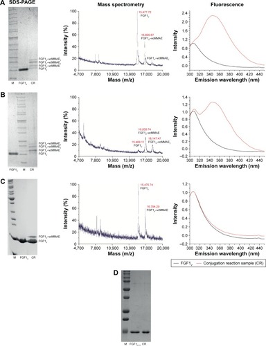
Figure 3 FGF1V–vcMMAE conjugate characteristics.
Notes: (A) Normalized thermal denaturation curve of FGF1V–vcMMAE conjugate and unconjugated FGF1V monitored by ellipticity changes. (B) In vitro cathepsin B cleavage of FGF1V–vcMMAE conjugate. FGF1V–vcMMAE was digested with cathepsin B at 37°C, pH 5.2 and 1:200 protease-to-FGF1 ratio. (C) Biological activity of FGF1V–vcMMAE and unconjugated FGF1V. Activation of signaling cascades in NIH 3T3 cells after incubation with increasing concentrations of FGF1V or FGF1V–vcMMAE detected with Western blot analysis. FGF1V–vcMMAE activation of FGFR was quantified to be 80% of unconjugated FGF1V at 10 ng/mL.
Abbreviations: FGF1V, fibroblast growth factor 1 variant; FGFR, fibroblast growth factor receptor; vcMMAE, valine–citrulline monomethyl auristatin E.
Abbreviations: FGF1V, fibroblast growth factor 1 variant; FGFR, fibroblast growth factor receptor; vcMMAE, valine–citrulline monomethyl auristatin E.
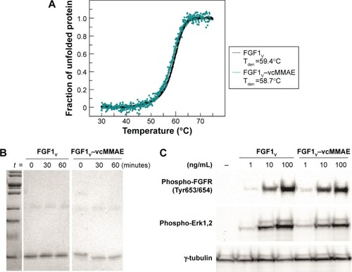
Figure 4 Viability of cells expressing FGFR treated with FGF1V–vcMMAE.
Notes: (A) FGFR1 expression in used cell lines. U20S FGFR1, U20S, MDA-MB-134-VI, and BJ cell lysates were analyzed by Western blot with anti-FGFR1 antibody; equal loading was confirmed by anti-γ-tubulin antibodies. (B) Viability of BJ, MDA-MB-134-VI, U20S FGFR1, and U20S cells after 96 hours of treatment with FGF1V, FGF1V–vcMMAE, or vcMMAE, in the presence of heparin, assessed with alamarBlue assay. Results shown are mean values from three experiments. Error bars indicate standard deviation.
Abbreviations: FGF1V, fibroblast growth factor 1 variant; FGFR, fibroblast growth factor receptor; vcMMAE, valine–citrulline monomethyl auristatin E.
Abbreviations: FGF1V, fibroblast growth factor 1 variant; FGFR, fibroblast growth factor receptor; vcMMAE, valine–citrulline monomethyl auristatin E.
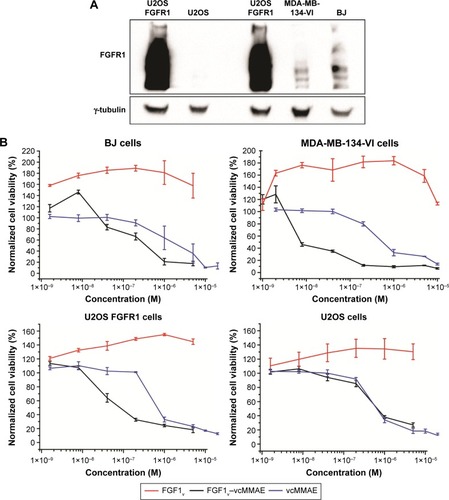
Table 1 Comparison of cytotoxic effects of FGF1V–vcMMAE conjugate and free vcMMAE
Figure 5 Proposed mechanism of action of FGF1V–vcMMAE conjugate.
Notes: After binding to the high affinity FGFRs on the cancer cell surface, FGF1V–vcMMAE conjugates are internalized and the cytotoxic drug is released from the conjugate either by linker cleavage in the endosomal compartment or after FGF1 degradation in the lysosome. The fully active cytotoxic drug leaves the lysosomal compartment and exerts its toxic action inhibiting microtubule polymerization and thereby leading to cell death.
Abbreviations: FGF1V, fibroblast growth factor 1 variant; FGFR, fibroblast growth factor receptor; MMAE, monomethyl auristatin E; vcMMAE, valine–citrulline monomethyl auristatin E.
Abbreviations: FGF1V, fibroblast growth factor 1 variant; FGFR, fibroblast growth factor receptor; MMAE, monomethyl auristatin E; vcMMAE, valine–citrulline monomethyl auristatin E.
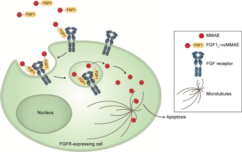
Figure S1 FGFRs mRNA expression levels in studied cell lines.
Notes: Relative expression level of different FGFRs were normalized by the ΔΔCT method to HPRT1 expression level, and shown as an expression level relative to human fibroblast cells (BJ). Experiments were performed in duplicates.
Abbreviations: FGFR, fibroblast growth factor receptor; mRNA, messenger RNA.
Abbreviations: FGFR, fibroblast growth factor receptor; mRNA, messenger RNA.
Figure S2 Time-dependency of FGF1V–vcMMAE cytotoxicity.
Notes: Viability of BJ cells after 24, 48, 72, and 96 hours of treatment with FGF1V, FGF1V–vcMMAE, or vcMMAE, in the presence of heparin, assessed with alamarBlue assay, n=2.
Abbreviations: FGF1V, fibroblast growth factor 1 variant; vcMMAE, valine–citrulline monomethyl auristatin E.
Abbreviations: FGF1V, fibroblast growth factor 1 variant; vcMMAE, valine–citrulline monomethyl auristatin E.
Figure S3 Apoptosis and cell death induced by FGF1V–vcMMAE.
Note: Flow cytometry analysis of U2OS, U2OS FGFR1, and MDA-MB-134-VI cells after the treatment with 6 µM FGF1V–vcMMAE and staining with annexin V-FITC and propidium iodide; experiments performed in duplicate.
Abbreviations: FGF1V, fibroblast growth factor 1 variant; FGFR, fibroblast growth factor receptor; vcMMAE, valine–citrulline monomethyl auristatin E.
Abbreviations: FGF1V, fibroblast growth factor 1 variant; FGFR, fibroblast growth factor receptor; vcMMAE, valine–citrulline monomethyl auristatin E.
Figure S4 Representative images of MDA-MB-134-VI cells after 96 hours of incubation with different concentrations of FGF1V, FGF1V–vcMMAE, or vcMMAE.
Abbreviations: FGF1V, fibroblast growth factor 1 variant; vcMMAE, valine–citrulline monomethyl auristatin E.