Figures & data
Figure 1 Scanning electron microscopy of mesalamine-coated microparticles.
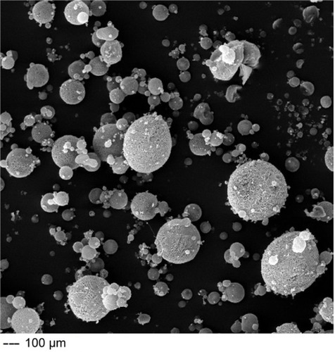
Figure 2 In vitro release profiles of different mesalamine formulations.
Abbreviations: PBS, phosphate-buffered saline; SD, standard deviation.
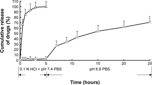
Table 1 Pharmacokinetic parameters of mesalamine after oral administration of mesalamine-coated microparticles and mesalamine suspensions to rats (n=6)
Figure 3 Plasma concentration–time profiles of mesalamine in rats after oral administration of mesalamine-coated microparticles and mesalamine suspensions (n=6).
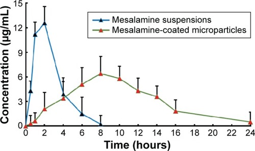
Table 2 The AUC0–24 h of mesalamine in stomach, small intestine, and colon after intragastric administration of coated microparticles and suspensions to mice (n=6)
Figure 4 Distribution of drugs in tissues of mice following intragastric administration of a single 10 mg/kg dose of mesalamine-coated microparticles and mesalamine suspensions.
Abbreviation: SD, standard deviation.
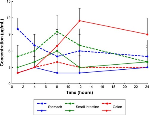
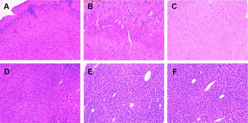
Figure 5 The images of hematoxylin and eosin staining tissue sections of mice after intragastric administration of mesalamine-coated microparticles and mesalamine suspensions.