Figures & data
Table 1 Effect of sitagliptin on BW, HW, and blood glucose in control and diabetic rats
Figure 1 Effect of sitagliptin on (A) troponin I level and (B) CK-MB activity.
Abbreviations: CK-MB, creatine kinase MB; SEM, standard error of the mean.
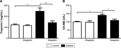
Figure 2 Effect of sitagliptin on serum lipid profile and cardiovascular indices.
Abbreviations: HDL, high-density lipoprotein; LDL, low-density lipoprotein; SEM, standard error of the mean; T cholesterol, total cholesterol; vLDL, very low-density lipoprotein.
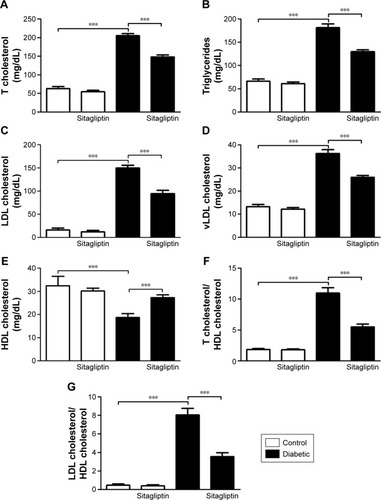
Figure 3 Effect of sitagliptin on oxidative stress and antioxidant defense system parameters.
Abbreviations: CAT, catalase; GSH, reduced glutathione; MDA, malondialdehyde; NO, nitric oxide; SEM, standard error of the mean; SOD, superoxide dismutase.
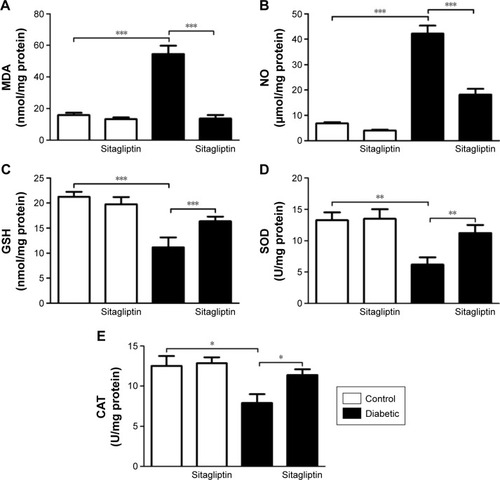
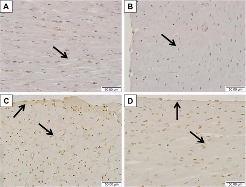
Figure 4 Effect of sitagliptin on cardiac IL-6 levels.
Abbreviations: IL-6, interleukin-6; SEM, standard error of the mean.
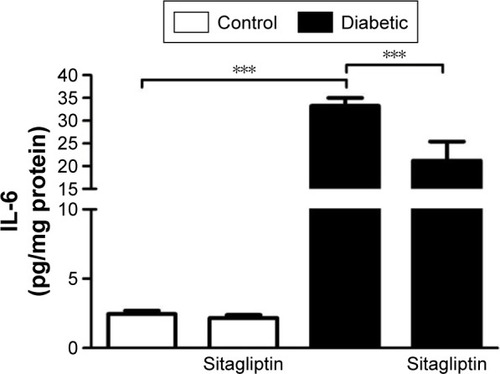
Figure 5 Effect of sitagliptin on histological changes in the heart of control and diabetic rats.
Abbreviation: H&E, hematoxylin and eosin.
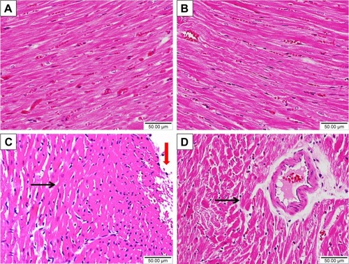
Figure 6 Effect of sitagliptin on collagen deposition in the heart of control and diabetic rats.
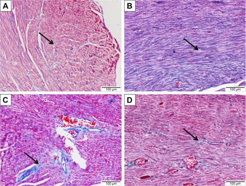
Figure 7 Effect of sitagliptin on JAK2 phosphorylation in the heart of control and diabetic rats.
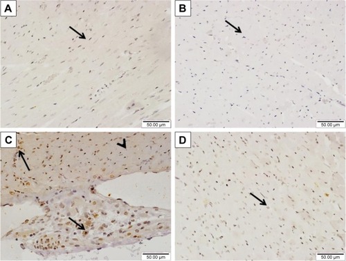
Figure 8 Effect of sitagliptin on STAT3 phosphorylation in the heart of control and diabetic rats.