Figures & data
Figure 1 Chemical structure of UA.
Abbreviation: UA, ursolic acid.
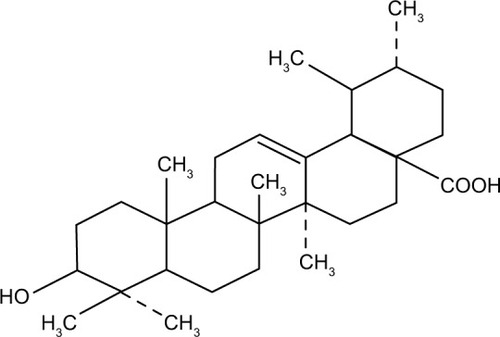
Figure 2 Nrf2 was overexpressed in cisplatin-resistant human hepatocellular carcinoma HepG2/DDP cells.
Notes: (A) HepG2 cells were treated with series concentration of cisplatin (0.1–25.6 μg/mL) for 48 hours. (B) HepG2/DDP cells were treated with series concentration of cisplatin (2–512 μg/mL) for 48 hours. (C) The level of Nrf2 and its downstream target genes HO-1, NQO1, and GST in HepG2 and HepG2/DDP cells was detected by Western blot assay. Results are representative of three different experiments, and they are expressed as mean ± SD.
Abbreviations: GAPDH, glyceraldehyde-3-phosphate dehydrogenase; GST, glutathione S-transferase; HepG2, hepatocellular carcinoma cell line; HepG2/DDP, cisplatin-resistant hepatocellular carcinoma cell line; HO-1, heme oxygenase-1; NQO1, NAD(P)H quinone oxidoreductase 1; Nrf2, nuclear factor erythroid-2-related factor 2; SD, standard deviation.
Abbreviations: GAPDH, glyceraldehyde-3-phosphate dehydrogenase; GST, glutathione S-transferase; HepG2, hepatocellular carcinoma cell line; HepG2/DDP, cisplatin-resistant hepatocellular carcinoma cell line; HO-1, heme oxygenase-1; NQO1, NAD(P)H quinone oxidoreductase 1; Nrf2, nuclear factor erythroid-2-related factor 2; SD, standard deviation.
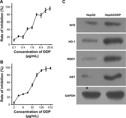
Figure 3 UA potentiates cisplatin-induced growth inhibition.
Notes: (A) HepG2 cells and HepG2/DDP cells were treated with increasing concentrations of UA (1.125–288 μg/mL) for 48 hours. (B) HepG2 cells were treated with series concentration of cisplatin (0.1–25.6 μg/mL) and UA (1.125 μg/mL and 2.25 μg/mL) for 48 hours. (C) HepG2/DDP cells were treated with series concentration of cisplatin (2–512 μg/mL) and UA (1.125 μg/mL and 2.25 μg/mL) for 48 hours. (D) HepG2 cells were treated for 48 hours with series concentration of cisplatin (0.1–25.6 μg/mL) and/or UA (1.125 μg/mL and 2.25 μg/mL). HepG2/DDP cells were incubated for 48 hours with series concentration of cisplatin (2–512 μg/mL) and/or UA (1.125 μg/mL and 2.25 μg/mL). Then, IC50 of cisplatin was calculated. *P<0.05 compared the HepG2/DDP cells alone treated with cisplatin. #P<0.05 vs the HepG2/DDP cells cotreated with cisplatin (2–512 μg/mL) and UA (1.125 μg/mL). Results are representative of three different experiments, and they are expressed as mean ± SD. The statistical analysis was performed using either one-way analysis of variance or two-tailed Student’s t-test for multiple comparisons.
Abbreviations: HepG2, hepatocellular carcinoma cell line; HepG2/DDP, cisplatin-resistant hepatocellular carcinoma cell line; IC50, 50% inhibitory concentration; SD, standard deviation; UA, ursolic acid.
Abbreviations: HepG2, hepatocellular carcinoma cell line; HepG2/DDP, cisplatin-resistant hepatocellular carcinoma cell line; IC50, 50% inhibitory concentration; SD, standard deviation; UA, ursolic acid.
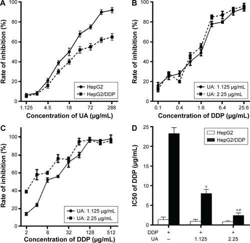
Figure 4 UA–cisplatin combination enhances low-dose cisplatin-induced apoptosis and causes G0/G1 arrest in resistant cells.
Notes: Analysis of cell apoptosis (A) and cell cycle (B) of HepG2/DDP cells treated with 8.92 μg/mL cisplatin (IC30 of cisplatin for HepG2/DDP cells) and/or UA (2.25 μg/mL) for 48 hours. *P<0.05, **P<0.01 compared to the cisplatin-alone-treated group. Results are representative of three different experiments, and they are expressed as mean ± SD. The statistical analysis was performed using either one-way analysis of variance or two-tailed Student’s t-test for multiple comparisons.
Abbreviations: HepG2/DDP, cisplatin–resistant hepatocellular carcinoma cell line; IC30, 30% inhibitory concentration; PI, propidium iodide; SD, standard deviation; UA, ursolic acid.
Abbreviations: HepG2/DDP, cisplatin–resistant hepatocellular carcinoma cell line; IC30, 30% inhibitory concentration; PI, propidium iodide; SD, standard deviation; UA, ursolic acid.
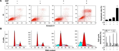
Figure 5 UA–cisplatin combination increases low-dose cisplatin-induced mitochondrial oxidative stress in resistant cells.
Notes: The level of ROS (A) and MMP (B) of HepG2/DDP cells treated with 8.92 μg/mL cisplatin and/or UA (2.25 μg/mL) for 48 hours was detected. **P<0.01 compared to the cisplatin-alone-treated group. Results are representative of three different experiments, and they are expressed as mean ± SD.
Abbreviations: DCFH-DA, dichloro-dihydro-fluorescein diacetate; HepG2/DDP, cisplatin-resistant hepatocellular carcinoma cell line; MMP, mitochondrial membrane potential; ROS, reactive oxygen species; SD, standard deviation; UA, ursolic acid.
Abbreviations: DCFH-DA, dichloro-dihydro-fluorescein diacetate; HepG2/DDP, cisplatin-resistant hepatocellular carcinoma cell line; MMP, mitochondrial membrane potential; ROS, reactive oxygen species; SD, standard deviation; UA, ursolic acid.
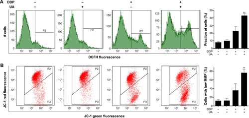
Figure 6 UA–cisplatin combination downregulates Nrf2 and its substrates.
Notes: The protein expression levels of Nrf2, HO-1, NQO1, and GST of HepG2/DDP cells treated with 8.92 μg/mL cisplatin and/or UA (2.25 μg/mL) for 48 hours were detected by Western blot analysis. Results are representative of three different experiments, and they are expressed as mean ± SD.
Abbreviations: GAPDH, glyceraldehyde-3-phosphate dehydrogenase; GST, glutathione S-transferase; HepG2/DDP, cisplatin-resistant hepatocellular carcinoma cell line; HO-1, heme oxygenase-1; NQO1, NAD(P)H quinone oxidoreductase 1; Nrf2, nuclear factor erythroid-2-related factor 2; SD, standard deviation; UA, ursolic acid.
Abbreviations: GAPDH, glyceraldehyde-3-phosphate dehydrogenase; GST, glutathione S-transferase; HepG2/DDP, cisplatin-resistant hepatocellular carcinoma cell line; HO-1, heme oxygenase-1; NQO1, NAD(P)H quinone oxidoreductase 1; Nrf2, nuclear factor erythroid-2-related factor 2; SD, standard deviation; UA, ursolic acid.
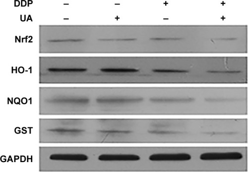
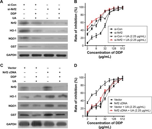
Figure 7 UA sensitizes HepG2/DDP cells to low-dose cisplatin via inhibition of Nrf2/ARE signaling pathway.
Notes: (A) HepG2/DDP cells were transfected with Nrf2 siRNA (si-Nrf2) or negative control (si-Con), or (C) HepG2/DDP cells were transfected with Nrf2 cDNA or empty vector (Vector), then treated with 8.92 μg/mL cisplatin (IC30 of cisplatin for HepG2/DDP cells) and/or UA (2.25 μg/mL) for 48 hours. The level of Nrf2, HO-1, NQO1, and GST was detected by Western blot analysis. (B) HepG2/DDP cells were transfected with si-Nrf2 or si-Con, or (D) HepG2/DDP cells were transfected with Nrf2 cDNA or empty vector (Vector), then treated with series concentration of cisplatin (2–512 μg/mL) and/or UA (2.25 μg/mL) for 48 hours. The inhibition rate of cell was detected by CCK8 assay. Results are representative of three different experiments, and they are expressed as mean ± SD.
Abbreviations: ARE, antioxidant response element; CCK8, Cell Counting Kit 8; cDNA, complementary DNA; GAPDH, glyceraldehyde-3-phosphate dehydrogenase; GST, glutathione S-transferase; HepG2/DDP, cisplatin–resistant hepatocellular carcinoma cell line; HO-1, heme oxygenase-1; IC30, 30% inhibitory concentration; NQO1, NAD(P)H quinone oxidoreductase 1; Nrf2, nuclear factor erythroid-2-related factor 2; SD, standard deviation; siRNA, small interfering RNA; UA, ursolic acid.
Abbreviations: ARE, antioxidant response element; CCK8, Cell Counting Kit 8; cDNA, complementary DNA; GAPDH, glyceraldehyde-3-phosphate dehydrogenase; GST, glutathione S-transferase; HepG2/DDP, cisplatin–resistant hepatocellular carcinoma cell line; HO-1, heme oxygenase-1; IC30, 30% inhibitory concentration; NQO1, NAD(P)H quinone oxidoreductase 1; Nrf2, nuclear factor erythroid-2-related factor 2; SD, standard deviation; siRNA, small interfering RNA; UA, ursolic acid.