
Figures & data
Table 1 Study scheme
Table 2 Body mass of rats
Figure 1 Morphology of the rat dorsolateral prostate.
Abbreviation: H&E, hematoxylin and eosin.
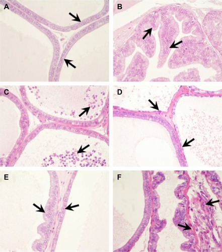
Figure 2 Ultrastructure of the rat dorsolateral prostate.
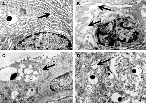
Figure 3 Immunolocalization and immunoexpression of PCNA (A, B), cytokeratin (C, D), and desmin (E, F) in the rat dorsolateral prostate.
Abbreviation: PCNA, proliferating cell nuclear antigen.
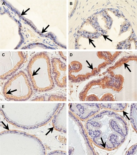
Figure 4 Percentage of PCNA-positive cells in epithelium and stroma in the rat dorsolateral prostate in control (I) and experimental groups (II–VIII).
Abbreviations: CsA, cyclosporin A; MMF, mycophenolate mofetil; Rapa, rapamycin; PCNA, proliferating cell nuclear antigen; Pred, prednisone; Tac, tacrolimus.
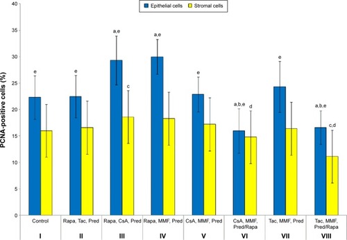
Figure 5 Percentage of cytokeratin-positive cells in epithelium in the rat dorsolateral prostate in control (I) and experimental groups (II–VIII).
Abbreviations: CsA, cyclosporin A; MMF, mycophenolate mofetil; Rapa, rapamycin; Pred, prednisone; Tac, tacrolimus.
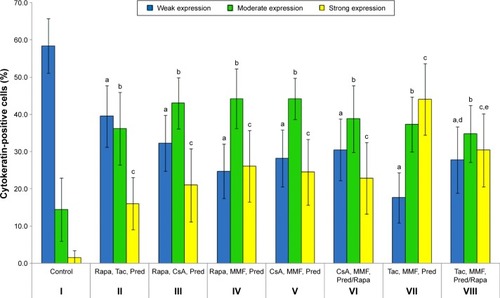
Figure 6 Percentage of desmin-positive cells in epithelium in the rat dorsolateral prostate in control (I) and experimental groups (II–VIII).
Abbreviations: CsA, cyclosporin A; MMF, mycophenolate mofetil; Rapa, rapamycin; Pred, prednisone; Tac, tacrolimus.
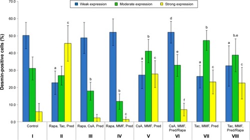
Table S1 Percentage of PCNA-positive epithelial and stromal cells in the rat dorsolateral prostate in control (I) and experimental (II–VIII) groups
Table S2 Percentage of cytokeratin-positive cells in the rat dorsolateral prostate in control (I) and experimental (II–VIII) groups
Table S3 Percentage of desmin-positive cells in the rat dorsolateral prostate in control (I) and experimental (II–VIII) groups