Figures & data
Figure 1 Structure of (A) quercetin and (B) isoquercitrin.
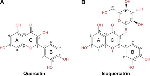
Figure 2 Structure of serine/threonine kinases.
Abbreviations: C-lobe, C-terminal lobe; DFG, Asp–Phe–Gly; N-lobe, N-terminal lobe; PDB, Protein Data Bank.
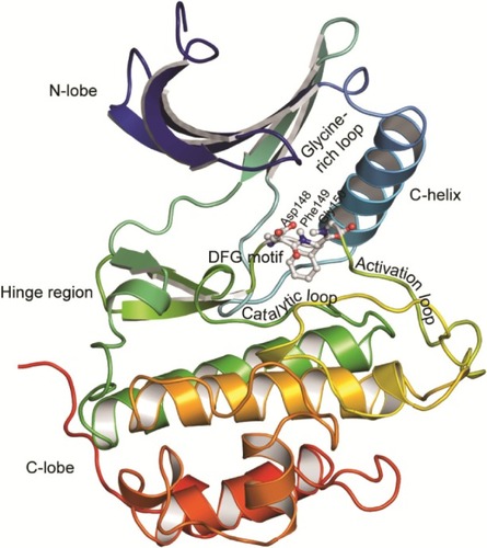
Table 1 Proteins and PDB IDs of structures used in this study
Table 2 Interactions of quercetin, isoquercitrin, and selected inhibitors with serine/threonine kinases
Figure 3 Docked pose and hydrogen bond interactions of quercetin and isoquercitrin with serine/threonine protein kinases.
Abbreviation: PDB, Protein Data Bank.
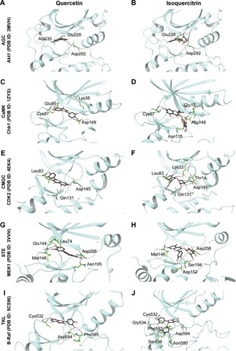
Figure S1 Docked pose and hydrogen bond interactions of quercetin and isoquercitrin with AGC family serine/threonine kinases.
Abbreviation: PDB, Protein Data Bank.
Figure S2 Docked pose and hydrogen bond interactions of quercetin and isoquercitrin with CMGC family serine/threonine kinases.
Abbreviation: PDB, Protein Data Bank.
Figure S3 Docked pose and hydrogen bond interactions of quercetin and isoquercitrin with STE family serine/threonine kinases.
Abbreviation: PDB, Protein Data Bank.