Figures & data
Figure 1 QuantiTect Primer Assay specification.
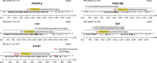
Figure 2 Time- and dose-dependent antiproliferation effect of imatinib mesylate with clinically relevant concentrations (2–10 µM) in two MCF7 and T-47D breast tumorigenic cell lines and a nontumorigenic breast cell line MCF 10A.
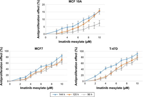
Figure 3 The concentration of imatinib mesylate that is required for 50% inhibition of two MCF7 and T-47D breast tumorigenic cell lines and a nontumorigenic breast cell line MCF 10A proliferation (half minimal inhibitory concentration) after 144 h treatment compared to untreated cell lines.
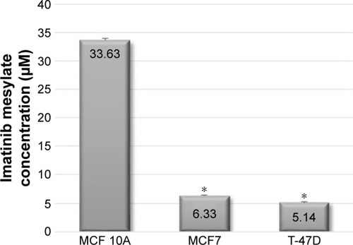
Figure 4 Apoptosis terminal deoxynucleotidyl transferase dUTP nick end labeling assay detection of time-dependent imatinib mesylate apoptosis induction within 48–144 h treatment with corresponding half minimal inhibitory concentration of imatinib mesylate for each cell line.
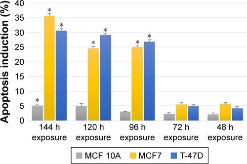
Figure 5 The effect of imatinib mesylate on cell cycle distribution of MCF7, T-47D tumorigenic and MCF 10A nontumorigenic breast cell lines.
Abbreviations: G, gap; M, mitosis; S, synthesis; PI-A, propidium iodide-A.
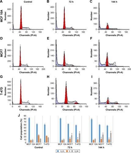
Figure 6 Comparative expression of the PDGFR-β, c-Kit, PDGF-BB and SCF genes compared to house-keeping gene (β-actin) in untreated two MCF7 and T-47D breast tumorigenic cell lines and a nontumorigenic breast cell line MCF 10A.
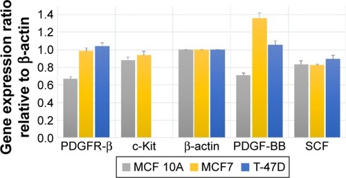
Figure 7 The relative gene expression pattern profiling of the PDGFR-β, c-Kit, PDGF-BB and SCF genes in imatinib mesylate-treated cell lines compared to untreated controls and normalized with HK genes.
Abbreviations: PDGF-BB, platelet-derived growth factor BB; PDGFR, platelet-derived growth factor receptor; SCF, stem cell factor.
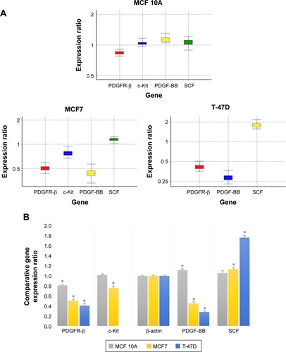
Table 1 The relative gene expression analysis report
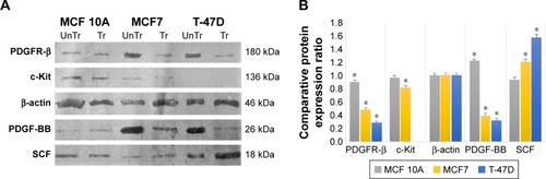
Figure 8 Relative expression of the PDGFR-β, c-Kit, PDGF-BB and SCF proteins compared to house-keeping protein (β-actin) in untreated cell lines.
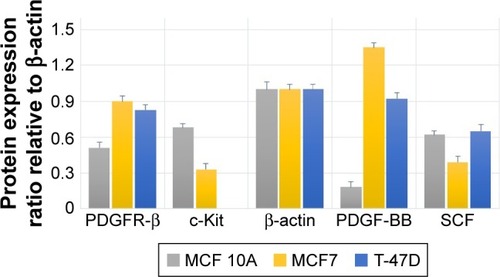
Figure 9 The effect of imatinib mesylate treatment on PDGFR-β, c-Kit, PDGF-BB and SCF protein expression related to untreated cell lines.
Abbreviations: PDGF-BB, platelet-derived growth factor BB; PDGFR, platelet-derived growth factor receptor; Tr, treated-cell lines; UnTr, untreated-cell lines.