Figures & data
Table 1 Oxidative and nitrosative stress parameters
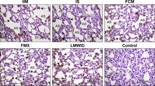
Figure 1 Nitrotyrosine immunohistochemistry.
Notes: Arrows indicate localization of nitrotyrosine immunostaining in lung of the different treatment groups. Positive staining (brown) is found in alveolar cells, primarily in groups treated with LMWID, FMX, and IIM. Cell types and tissue are indicated as capillary endothelial cells (CEC), alveolar sac (AS), alveolar cells (AC), and interalveolar septum (IAS). Original magnification ×200. Scale bar 50 μm. IIM, iron isomaltoside 1000 (Monofer®, Pharmacosmos A/S, Holbæk, Denmark); IS, iron sucrose (Venofer®, American Regent, Shirley, NY, USA); FCM, ferric carboxymaltose (Ferinject®, Vifor (International) Ltd., St Gallen, Switzerland); FMX, ferumoxytol (Feraheme®, AMAG Pharmaceuticals Inc., Lexington, MA, USA); LMWID, low molecular weight iron dextran (Infed®, Watson Pharma Inc., Morristown, NJ, USA); control, saline treatment.
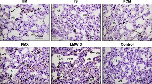
Figure 2 Nitrotyrosine Western blot analysis of lung homogenates.
Notes: (A) Nitrotyrosine immunoreactivity by Western blot technique for the different treatment groups. Arrows denote tyrosine nitration for proteins in the range of approximately 65 kDa and 58 kDa. (B) Densitometric quantitation of tyrosine nitration for proteins resolved by Western blot shown in (A). Bars (with SD) represent total amount of tyrosine nitrated proteins as quantified by densitometry. Tyrosine nitration was significantly (P<0.01) greater in LMWID-, FMX-, and IIM-treated rats compared to control, whereas this parameter was not different between FCM and IS treatment compared to controls. Tyrosine nitration was also significantly (P<0.01) increased in the LMWID and FMX groups compared to all other intravenous iron groups. IIM, iron isomaltoside 1000 (Monofer®, Pharmacosmos A/S, Holbæk, Denmark); IS, iron sucrose (Venofer®, American Regent, Shirley, NY, USA); FCM, ferric carboxymaltose (Ferinject®, Vifor (International) Ltd., St Gallen, Switzerland); FMX, ferumoxytol (Feraheme®, AMAG Pharmaceuticals Inc., Lexington, MA, USA); LMWID, low molecular weight iron dextran (Infed®, Watson Pharma Inc., Morristown, NJ, USA); control, saline treatment.
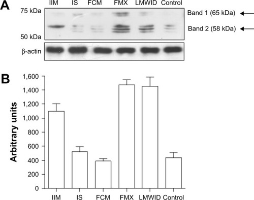
Table 2 Inflammation parameters
Figure 3 HIF-1α immunohistochemistry.
Notes: Arrows indicate localization of HIF-1α immunostaining in alveolar cells in lung of the different treatment groups. Positive staining (brown) is found in alveolar cells, interalveolar septum, and capillary endothelial cells, predominantly in the LMWID, FMX, and IIM treatment groups. Cell types and tissue are indicated as capillary endothelial cells (CEC), alveolar sac (AS), alveolar cells (AC), and interalveolar septum (IAS). Original magnification ×200. Scale bar 50 μm. IIM, iron isomaltoside 1000 (Monofer®, Pharmacosmos A/S, Holbæk, Denmark); IS, iron sucrose (Venofer®, American Regent, Shirley, NY, USA); FCM, ferric carboxymaltose (Ferinject®, Vifor (International) Ltd., St Gallen, Switzerland); FMX, ferumoxytol (Feraheme®, AMAG Pharmaceuticals Inc., Lexington, MA, USA); LMWID, low molecular weight iron dextran (Infed®, Watson Pharma Inc., Morristown, NJ, USA); control, saline treatment.
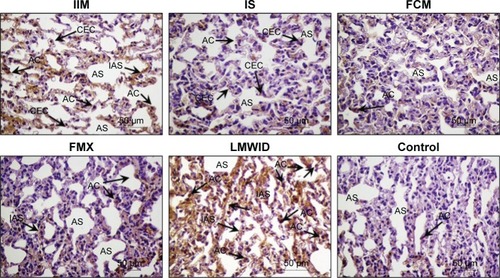
Figure 4 ED1 immunohistochemistry.
Notes: Arrows indicate presence of ED1 macrophages in lung interstitium in the different treatment groups. Numbers of positively stained (brown) cells (ED1 macrophages) are greatest in the LMWID, FMX, and IIM treatment groups. Cell types and tissue are indicated as alveolar sac (AS), and interstitial macrophages (IM). Original magnification ×200. Scale bar 50 μm. IIM, iron isomaltoside 1000 (Monofer®, Pharmacosmos A/S, Holbæk, Denmark); IS, iron sucrose (Venofer®, American Regent, Shirley, NY, USA); FCM, ferric carboxymaltose (Ferinject®, Vifor (International) Ltd., St Gallen, Switzerland); FMX, ferumoxytol (Feraheme®, AMAG Pharmaceuticals Inc., Lexington, MA, USA); LMWID, low molecular weight iron dextran (Infed®, Watson Pharma Inc., Morristown, NJ, USA); control, saline treatment.
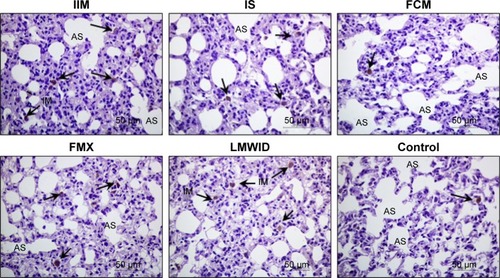
Table 3 Tissue iron and ferritin
Figure 5 Prussian blue staining.
Notes: Identification of iron deposits in lung at the end of study. Cell types and tissue are indicated as capillary endothelial cells (CEC), alveolar sac (AS), alveolar cells (AC), interstitial macrophages (IM), and interalveolar septum (IAS). Arrows denote positive staining for iron deposits (blue) predominantly in the group treated with LMWID (CEC, AC, IM, IAS), with moderate amounts in the FMX (CEC) and IIM (IAS) treatment groups. IS, FCM, and control groups show similar low level staining. Original magnification ×200. Scale bar 50 μm. IIM, iron isomaltoside 1000 (Monofer®, Pharmacosmos A/S, Holbæk, Denmark); IS, iron sucrose (Venofer®, American Regent, Shirley, NY, USA); FCM, ferric carboxymaltose (Ferinject®, Vifor (International) Ltd., St Gallen, Switzerland); FMX, ferumoxytol (Feraheme®, AMAG Pharmaceuticals Inc., Lexington, MA, USA); LMWID, low molecular weight iron dextran (Infed®, Watson Pharma Inc., Morristown, NJ, USA); control, saline treatment.
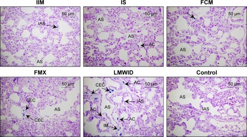
Figure S1 NF-κB65 immunohistochemistry.
Notes: Arrows indicate alveolar cells with positive staining for NF-κB. Original magnification ×200. Scale bar 50 μm. Cell types and tissue are indicated as alveolar sac (AS), and alveolar cells (AC). IIM, iron isomaltoside 1000 (Monofer®, Pharmacosmos A/S, Holbæk, Denmark); IS, iron sucrose (Venofer®, American Regent, Shirley, NY, USA); FCM, ferric carboxymaltose (Ferinject®, Vifor (International) Ltd., St Gallen, Switzerland); FMX, ferumoxytol (Feraheme®, AMAG Pharmaceuticals Inc., Lexington, MA, USA); LMWID, low molecular weight iron dextran (Infed®, Watson Pharma Inc., Morristown, NJ, USA); control, saline treatment.
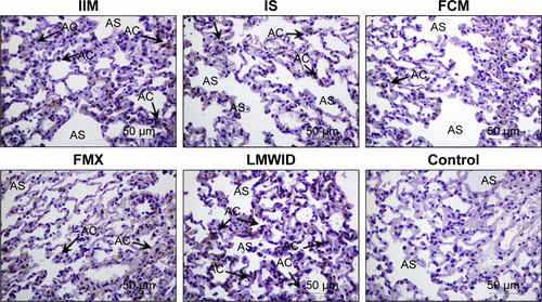
Figure S2 TNF-α immunohistochemistry.
Notes: Arrows indicate localization of TNF-α in alveolar cells. Original magnification ×200. Scale bar 50 μm. Cell types and tissue are indicated as alveolar sac (AS), and alveolar cells (AC). IIM, iron isomaltoside 1000 (Monofer®, Pharmacosmos A/S, Holbæk, Denmark); IS, iron sucrose (Venofer®, American Regent, Shirley, NY, USA); FCM, ferric carboxymaltose (Ferinject®, Vifor (International) Ltd., St Gallen, Switzerland); FMX, ferumoxytol (Feraheme®, AMAG Pharmaceuticals Inc., Lexington, MA, USA); LMWID, low molecular weight iron dextran (Infed®, Watson Pharma Inc., Morristown, NJ, USA); control, saline treatment.
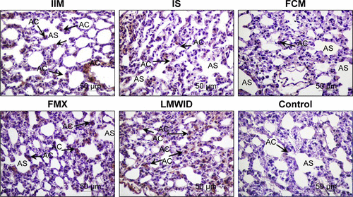
Figure S3 IL-6 immunohistochemistry.
Notes: Arrows indicate localization of IL-6 in alveolar cells. Original magnification ×200. Scale bar 50 μm. Cell types and tissue are indicated as alveolar sac (AS), and alveolar cells (AC). IIM, iron isomaltoside 1000 (Monofer®, Pharmacosmos A/S, Holbæk, Denmark); IS, iron sucrose (Venofer®, American Regent, Shirley, NY, USA); FCM, ferric carboxymaltose (Ferinject®, Vifor (International) Ltd., St Gallen, Switzerland); FMX, ferumoxytol (Feraheme®, AMAG Pharmaceuticals Inc., Lexington, MA, USA); LMWID, low molecular weight iron dextran (Infed®, Watson Pharma Inc., Morristown, NJ, USA); control, saline treatment.
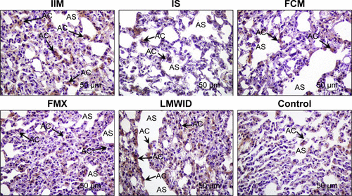
Figure S4 VEGF immunohistochemistry.
Notes: Arrows indicate presence of VEGF not only in endothelial cells’ localization but also in alveolar cells in the different groups. Original magnification ×200. Scale bar 50 μm. Cell types and tissue are indicated as alveolar sac (AS). IIM, iron isomaltoside 1000 (Monofer®, Pharmacosmos A/S, Holbæk, Denmark); IS, iron sucrose (Venofer®, American Regent, Shirley, NY, USA); FCM, ferric carboxymaltose (Ferinject®, Vifor (International) Ltd., St Gallen, Switzerland); FMX, ferumoxytol (Feraheme®, AMAG Pharmaceuticals Inc., Lexington, MA, USA); LMWID, low molecular weight iron dextran (Infed®, Watson Pharma Inc., Morristown, NJ, USA); control, saline treatment.
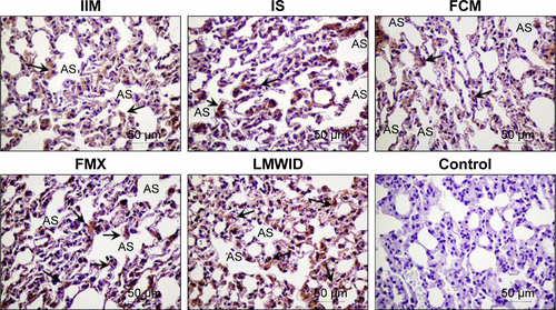
Figure S5 VCAM-1 immunohistochemistry.
Notes: Arrows indicate presence of VCAM in endothelial cells, alveolar cells, and also in lung interstitium in the different groups. Original magnification ×200. Scale bar 50 μm. Cell types and tissue are indicated as alveolar sac (AS). IIM, iron isomaltoside 1000 (Monofer®, Pharmacosmos A/S, Holbæk, Denmark); IS, iron sucrose (Venofer®, American Regent, Shirley, NY, USA); FCM, ferric carboxymaltose (Ferinject®, Vifor (International) Ltd., St Gallen, Switzerland); FMX, ferumoxytol (Feraheme®, AMAG Pharmaceuticals Inc., Lexington, MA, USA); LMWID, low molecular weight iron dextran (Infed®, Watson Pharma Inc., Morristown, NJ, USA); control, saline treatment.
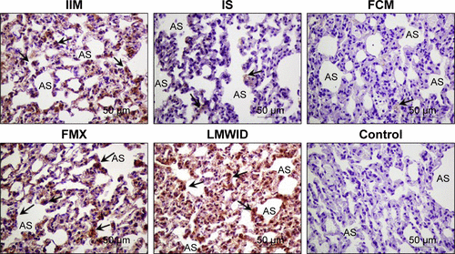
Figure S6 Ferritin L chain immunohistochemistry.
Notes: Arrows indicate localization of ferritin L in alveolar cells and other cells of the different groups. Original magnification ×200. Scale bar 50 μm. Cell types and tissue are indicated as alveolar sac (AS). IIM, iron isomaltoside 1000 (Monofer®, Pharmacosmos A/S, Holbæk, Denmark); IS, iron sucrose (Venofer®, American Regent, Shirley, NY, USA); FCM, ferric carboxymaltose (Ferinject®, Vifor (International) Ltd., St Gallen, Switzerland); FMX, ferumoxytol (Feraheme®, AMAG Pharmaceuticals Inc., Lexington, MA, USA); LMWID, low molecular weight iron dextran (Infed®, Watson Pharma Inc., Morristown, NJ, USA); control, saline treatment.
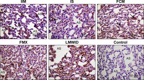
Figure S7 Ferritin H chain immunohistochemistry.
Notes: Arrows indicate localization of ferritin H in alveolar cells of the different groups. Original magnification ×200. Scale bar 50 μm. Cell types and tissue are indicated as alveolar sac (AS). IIM, iron isomaltoside 1000 (Monofer®, Pharmacosmos A/S, Holbæk, Denmark); IS, iron sucrose (Venofer®, American Regent, Shirley, NY, USA); FCM, ferric carboxymaltose (Ferinject®, Vifor (International) Ltd., St Gallen, Switzerland); FMX, ferumoxytol (Feraheme®, AMAG Pharmaceuticals Inc., Lexington, MA, USA); LMWID, low molecular weight iron dextran (Infed®, Watson Pharma Inc., Morristown, NJ, USA); control, saline treatment.