
Figures & data
Table 1 The mean serum level of bone resorption and formation markers among the five groups
Figure 1 Histopathological examination of rats’ bones of the five groups by light microscopy stained by H&E. (×200).
Notes: Sections with 3 µm thickness were prepared and stained with H&E. The cortical thickness and trabecular area in femur and vertebra were examined. Histopathological examination of rat bones showed numerous trabeculae, osteoblast cells and blood vessels in the atorvastatin group. These have been indicated by arrowheads and stars. (A) Group I: Section in a femur bone, revealed part of compact bone formed of osteons surrounded by several lamellae of bone, connected by Volkmann’s canal (arrow). (B) Group I: Section in a femur bone showed part of skeletal muscle fibers attached to the outer fibrous periosteum (arrow). Note the presence of osteons surrounded by interstitial lamellae (star). The inner circumferential lamellae (arrowhead) were separating the bone from the marrow cavity. (C) Group II: Section in a femur bone revealed large open spaces of bone marrow lined by endosteum (arrow) and separated by trabeculae of bone composed of several lamellae. (D) Group II: Section in a lumbar vertebra revealed large open spaces filled with bone marrow (star) and separated by trabeculae of well-formed bone (arrow) composed of several lamellae. (E) Group III: Section in a femur bone revealed a zone of calcifying cartilage (upper right). The rest of the slide showed numerous trabeculae of bone (star) with wide spaces of bone marrow, osteogenic cells and blood vessels. (F) Group III: Section in a femur bone revealed interlacing trabeculae of intensely basophilic trabeculae of woven bone with large and round osteocytes (star). The lower part of the section showed proliferated blood vessels, osteoclasts forming shallow depressions on the bone (arrow) and part of the periosteal tissue. (G) Group IV: Section in a femur bone revealed widely spaced thin bone trabeculae (arrow) lined by flattened osteoblasts, with fatty marrow filling the spaces between the trabeculae. (H) Group IV: Section in lumbar vertebra revealed widely spaced thin bone trabeculae, surrounding scant bone marrow material. (I) Group V: Section in a lumbar vertebral bone revealed trabeculae of bone formed of lenticular-shaped osteocytes (arrow) surrounding wide spaces filled with a scant amount of bone marrow (star). (J) Group V: Section in a femur showed trabeculae of bone surrounding wide spaces filled with a scant amount of bone marrow (star).
Abbreviation: H&E, hematoxylin and eosin.
Abbreviation: H&E, hematoxylin and eosin.
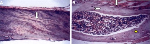
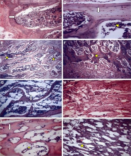
Table 2 Demographic data of the examined 70 postmenopausal patients
Table 3 T-scores of femoral neck, lumbar spine and distal radius of the examined 70 patients at the start and at the end of the 18 months study period