Figures & data
Figure 1 Andro synergistically enhanced the antitumor effects of 5-Fu on HCT-116 cells.
Notes: (A) HCT-116 cells were treated with 5-Fu ranging from 0 to 40 μM, respectively, for 24, 48, and 72 hours. The cell growth inhibition rate was measured by MTT assay. (B) The cells were treated with 5-Fu, Andro, or 5-Fu plus different concentrations of Andro, respectively, for 24, 48, and 72 hours. The cell growth inhibition rate was measured by MTT assay. (C) The cells were treated with 5-Fu, Andro, or Andro plus 5-Fu for 72 hours. Morphological changes were observed using a microscope at 200× magnification. (D) HCT-116 cells were treated with 5-Fu ranging from 0 to 16 μM combined with 10 μM Andro for 72 hours. Each experiment was repeated more than three times independently. (A, B, and D) Data are shown as mean ± SD, n=3, *p<0.05.
Abbreviations: Andro, andrographolide; MTT, 3-(4,5-dimethyl-2-thiazolyl)-2,5-diphenyl-2H-tetrazolium bromide; 5-Fu, 5-fluorouracil; SD, standard deviation; DMSO, dimethyl sulfoxide.
Abbreviations: Andro, andrographolide; MTT, 3-(4,5-dimethyl-2-thiazolyl)-2,5-diphenyl-2H-tetrazolium bromide; 5-Fu, 5-fluorouracil; SD, standard deviation; DMSO, dimethyl sulfoxide.
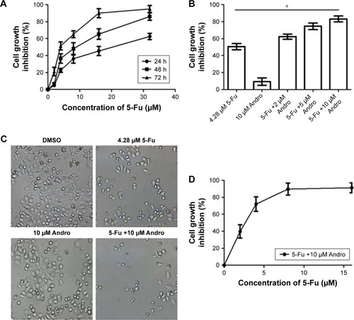
Table 1 IC50 and CI values for 5-Fu single dose (from 0 to 32 μM) or 5-Fu plus Andro (10 μM) in HCT-116 cells in 72 hours
Figure 2 5-Fu-induced apoptotic activities were enhanced by Andro against HCT-116 cells in vitro.
Notes: (A) HCT-116 cells were treated with 5-Fu, Andro, or Andro plus 5-Fu for 72 hours. Thereafter, the cells were collected and stained with Annexin V and PI. The apoptotic cells were observed by flow cytometry. (B) The levels of caspase-3 and cleaved caspase-3 were analyzed by Western blot analysis followed by the indicated treatment. Each experiment was repeated more than three times independently.
Abbreviations: Andro, andrographolide; 5-Fu, 5-fluorouracil; SD, standard deviation; PI, propidium iodide; DMSO, dimethyl sulfoxide; FITC, fluorescein isothiocyanate.
Abbreviations: Andro, andrographolide; 5-Fu, 5-fluorouracil; SD, standard deviation; PI, propidium iodide; DMSO, dimethyl sulfoxide; FITC, fluorescein isothiocyanate.
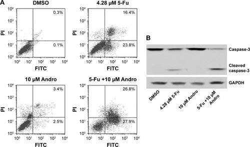
Figure 3 5-Fu-induced HCT-116 cells apoptosis was enhanced by Andro by inhibition of c-MET pathway in vitro.
Notes: (A) HCT-116 cells were treated with 5-Fu, Andro, or Andro plus 5-Fu for 72 hours. The expression levels of epidermal growth factors p-EGFR, p-FGFR2, p-VEGFR1, p-MET, p-HER2, p-ALK, and p-VEGFR2 were measured using ELISA kit. (B) The expression levels of c-MET, p-MET, p-AKT, and p-ERK were analyzed by Western blot analysis. (C) The HCT-116 cells were treated with 5-Fu, Andro plus 5-Fu, or three combinations for 72 hours. The cell growth inhibition rate was measured by MTT assay. Each experiment was repeated more than three times independently. (A and C) Data are shown as mean ± SD, n=3, *p<0.05.
Abbreviations: Andro, andrographolide; MTT, 3-(4,5-dimethyl-2-thiazolyl)-2,5-diphenyl-2H-tetrazolium bromide; 5-Fu, 5-fluorouracil; SD, standard deviation; DMSO, dimethyl sulfoxide; p-EGFR, phosphorylated epidermal growth factor receptor; p-FGFR2, phosphorylated fibroblast growth factor receptor 2; p-VEGFR1, phosphorylated vascular endothelial growth factor receptor 1; p-MET, phosphorylated cellular-mesenchymal to epithelial transition factor; p-HER2, phosphorylated human epidermal growth factor receptor 2; p-ALK, phosphorylated anaplastic lymphoma kinase; p-VEGFR2, phosphorylated vascular endothelial growth factor receptor 2; ELISA, enzyme linked immunosorbent assay; OD, optical density; HGF, hepatocyte growth factor.
Abbreviations: Andro, andrographolide; MTT, 3-(4,5-dimethyl-2-thiazolyl)-2,5-diphenyl-2H-tetrazolium bromide; 5-Fu, 5-fluorouracil; SD, standard deviation; DMSO, dimethyl sulfoxide; p-EGFR, phosphorylated epidermal growth factor receptor; p-FGFR2, phosphorylated fibroblast growth factor receptor 2; p-VEGFR1, phosphorylated vascular endothelial growth factor receptor 1; p-MET, phosphorylated cellular-mesenchymal to epithelial transition factor; p-HER2, phosphorylated human epidermal growth factor receptor 2; p-ALK, phosphorylated anaplastic lymphoma kinase; p-VEGFR2, phosphorylated vascular endothelial growth factor receptor 2; ELISA, enzyme linked immunosorbent assay; OD, optical density; HGF, hepatocyte growth factor.
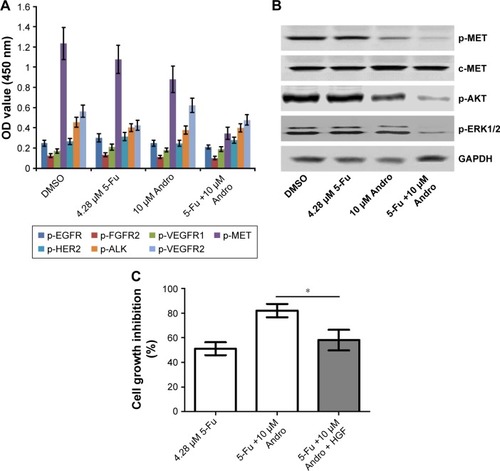
Figure 4 Andro enhanced the antitumor effect of 5-Fu on HCT-116 xenograft in vivo.
Notes: (A) HCT-116 tumor bearing mice were administered vehicle, 10 mg/kg 5-Fu (weekly, ip), 10 mg/kg Andro (daily, po), or the combination treatment of 5-Fu with Andro. Tumor volumes were monitored weekly. Data are shown as mean ± SD, n=6. (B) The tumors were collected after 4 weeks treatment and weighted.
Abbreviations: Andro, andrographolide; 5-Fu, 5-fluorouracil; SD, standard deviation; DMSO, dimethyl sulfoxide; ip, intraperitoneal; po, per oral.
Abbreviations: Andro, andrographolide; 5-Fu, 5-fluorouracil; SD, standard deviation; DMSO, dimethyl sulfoxide; ip, intraperitoneal; po, per oral.
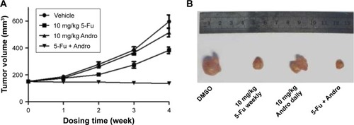
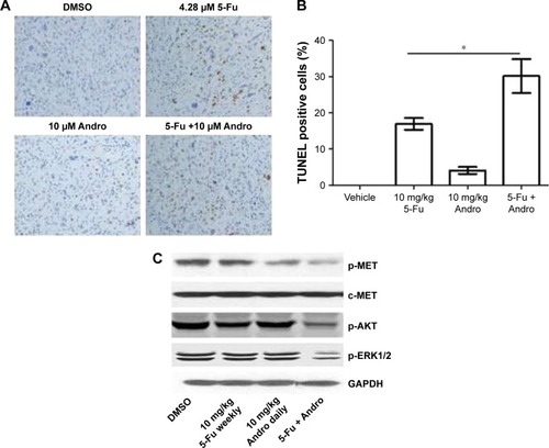
Figure 5 5-Fu-induced tumor apoptosis was enhanced by Andro due to p-MET level downregulation in vivo.
Notes: (A, B) The mice were sacrificed after 4 weeks of treatment; the tumors were removed and dewaxed. The apoptotic rate was measured using TUNEL assay and immunohistochemistry. (B) Data are shown as mean ± SD, n=3, *p<0.05. (C) The expression levels of c-MET, p-MET, p-AKT, and p-ERK were analyzed by Western blot in tumor tissue. Each experiment was repeated more than three times independently.
Abbreviations: Andro, andrographolide; 5-Fu, 5-fluorouracil; SD, standard deviation; DMSO, dimethyl sulfoxide; TUNEL, terminal dUTP nick end-labeling; p-MET, phosphorylated cellular-mesenchymal to epithelial transition factor.
Abbreviations: Andro, andrographolide; 5-Fu, 5-fluorouracil; SD, standard deviation; DMSO, dimethyl sulfoxide; TUNEL, terminal dUTP nick end-labeling; p-MET, phosphorylated cellular-mesenchymal to epithelial transition factor.