
Figures & data
Figure 1 Schematic of single probiotics nanoencapsulation and against harsh environment in human digestive system. (a) Representation of probiotics without nanocoating. (b) Nanoencapsulation of probiotics. (c) Gut bacteria pathway via the gastrointestinal tract and the importance of probiotics encapsulation. Data from these studies.Citation22–25
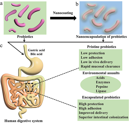
Figure 2 (a) Layer-by-layer self-assembly as the most widely utilized approach to the formation of polymeric nanocoating. After deposition of each layer, cells are collected by centrifugation, washed with buffer, and then collected again by centrifugation. (b) Schematic layer-by-layer templating of chitosan and alginate on bacteria surface.
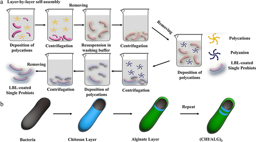
Figure 3 Schematic of probiotics-mediated oxidation of polyphenolic compounds bearing catechol groups by probiotic cells. (a) Structures of the phenolic precursors used in this study. (b) Process overview of probiotics-mediated oxidation of catechol compounds and in situ nanoencapsulation. (c) Possible mechanism of the probiotics-mediated phenolic oxidation using probiotics. Data from these studies.Citation37,Citation38
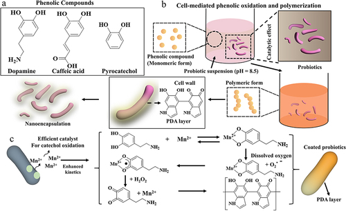
Figure 4 (a) Schematic representation of interfacial supramolecular self-assembly of FeIII and TA at various interfaces. FeIII ions are preloaded into one phase (Phase A), and the supramolecular self-assembly is induced at the interface of two immiscible phases by TA in the other phase (Phase B). (b) Schematic representation for formation of FeIII–TA-MOC hollow nanoshell. (c) Schematic representation for self-responsive FeIII-TA-MOC nanoshell formation on baker’s yeast.
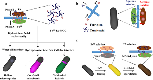
Figure 5 (a) Schematic illustration for the preparation of EM-coated-EcN by extruding bacteria with cell membranes. (b) Representative transmission electron microscope images of uncoated EcN and EM-coated-EcN. (c) A representative image of membrane shedding captured by TEM. (d) Schematic illustration for the removal of coating membranes. Adapted from Cao ZP, Cheng SS, Wang XY et al. Camouflaging bacteria by wrapping with cell membranes. Nat Commun. 2019. 10 (1): 3452. Creative Commons.Citation60
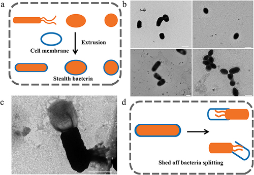
Figure 6 (a) Schematic illustration of the preparation of lipid membrane coated bacteria by biointerfacial supramolecular self-assembly. (b) The presence of coating membranes endows probiotic bacteria with exceptional resistance to various harsh environmental conditions. Adapted from Cao ZP, Wang XY, Pang Y et al. Biointerfacial self-assembly generates lipid membrane coated bacteria for enhanced oral delivery and treatment. Nat Commun. 2019. 10 (1): 5783. Creative Commons.Citation63
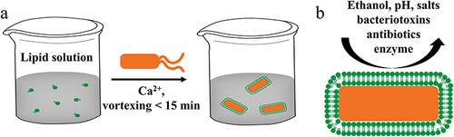
Table 1 The Advantages and Disadvantages of Different Nanoshell to Protect Probiotics