Figures & data
Figure 1 Chemical structure and purity of BM.
Abbreviations: BM, β-mangostin; HPLC, high-performance liquid chromatography; min, minutes; PDA, photodiode array; mAU, milli absorption unit.
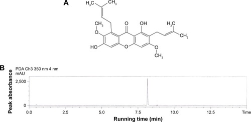
Table 1 Effects of BM administered intraduodenally on the biochemical parameters of gastric juice obtained from pylorus ligature in rats
Table 2 Observed ulcer area and inhibition percentage of BM against ethanol-induced gastric ulcer in rats
Figure 2 Macroscopic evaluation.
Abbreviation: BM, β-mangostin.
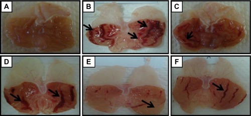
Table 3 Serum biochemical analysis of animal pretreated by BM against ethanol-induced gastric ulcer
Table 4 Lesion score of rat gastric tissue pretreated with BM against ethanol-induced gastric damage
Figure 3 Histological evaluation.
Abbreviations: BM, β-mangostin; H&E, hematoxylin and eosin.
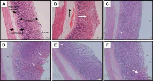
Figure 4 PAS staining.
Abbreviations: BM, β-mangostin; PAS, periodic acid–Schiff.
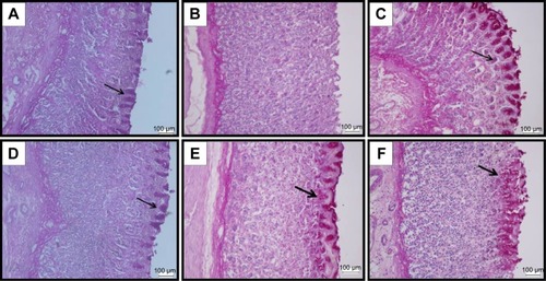
Figure 5 Effects of BM (BM; 5, 10, and 20 mg/kg bw) and omeprazole (OMP; 20 mg/kg bw) on (A) gastric mucosal PGE2 levels and (B) alcian blue binding to free gastric mucus against ethanol-induced gastric ulcer in rat.
Abbreviations: ANOVA, analysis of variance; BM, β-mangostin; OMP, omeprazole; PGE2, prostaglandin E2; SEM, standard error of the mean; bw, body weight.
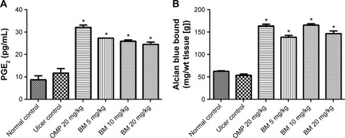
Figure 6 Effects of BM (5, 10, and 20 mg/kg bw) and OMP (20 mg/kg bw) on (A) GSH levels, (B) MDA level, (C) SOD activity, and (D) CAT activity against ethanol-induced gastric ulcer in rat.
Abbreviations: ANOVA, analysis of variance; BM, β-mangostin; CAT, catalase; GSH, glutathione; MDA, malondialdehyde; OMP, omeprazole; SEM, standard error of the mean; SOD, superoxide dismutase; bw, body weight.
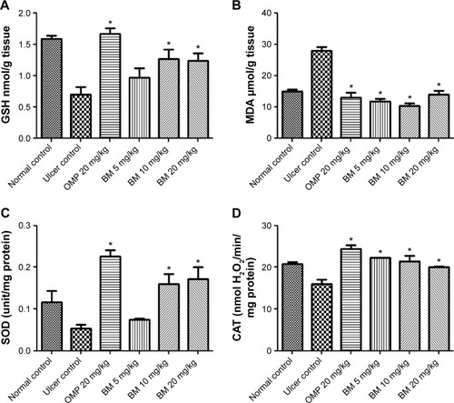
Figure 7 Effects of BM (5, 10, and 20 mg/kg bw) and OMP (20 mg/kg bw) on gastric mucosal NP-SH content against ethanol-induced gastric ulcer in rat.
Abbreviations: ANOVA, analysis of variance; BM, β-mangostin; NP-SH, nonprotein sulfhydryls; OMP, omeprazole; SEM, standard error of the mean; bw, body weight.
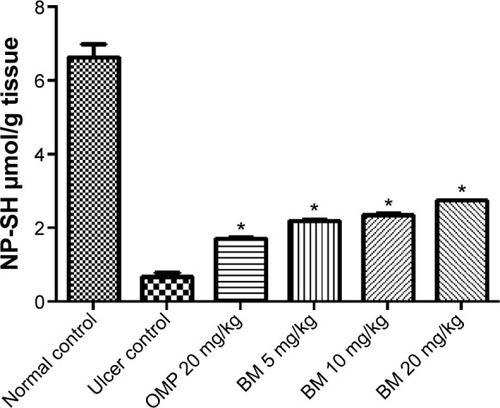
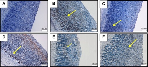
Figure 8 HSP70 IHC.
Abbreviations: BM, β-mangostin; HSP70, heat shock protein 70; IHC, immunohistochemistry.
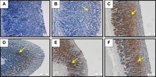
Figure 9 Bax IHC.
Abbreviations: BM, β-mangostin; IHC, immunohistochemistry.