Figures & data
Figure 1 Inhibitory effects of YSV on adhesion to Matrigel of human lung cancer cells in vitro.
Notes: Cells were pretreated with different concentrations of YSV (0.1 mg/mL, 0.2 mg/mL, 0.4 mg/mL, 0.8 mg/mL) for 24 hours, 48 hours, and 72 hours. Cell suspensions were placed into the wells of a 96-well culture plate coated with Matrigel and incubated for 1 hour at 37°C. The OD value of cells was measured by MTS assay. Values represent the mean ± SD. Bars indicate SD. *P<0.05, compared to control group, tested using one-way ANOVA and Student–Newman–Keuls test (N=8). (A) 95D: OD value. (B) 95D: inhibition ratio. (C) A549: OD value. (D) A549: inhibition ratio. (E) NCI-H1299: OD value. (F) NCI-H1299: inhibition ratio.
Abbreviations: YSV, tyroservatide; OD, optical density; SD, standard deviation; ANOVA, analysis of variance.
Abbreviations: YSV, tyroservatide; OD, optical density; SD, standard deviation; ANOVA, analysis of variance.
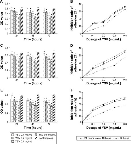
Figure 2 Inhibitory effects of YSV on invasion of human lung cancer cells in vitro.
Notes: The 95D, A549, and NCI-H1299 cells were pretreated with different concentrations of YSV (0.2 mg/mL, 0.4 mg/mL) for 48 hours. Then, 100 μL/well cell suspension (2×10⁶/mL) was placed into the upper chambers of the wells. The cells that had migrated to the lower surface of the filter were stained with hematoxylin and eosin and counted under a microscope. Values represent the mean ± SD. Bars indicate SD. (A) 95D. (B) A549. (C) NCI-H1299. *P<0.05, compared to control group, tested using one-way ANOVA and Student–Newman–Keuls test (N=3).
Abbreviations: YSV, tyroservatide; SD, standard deviation; ANOVA, analysis of variance.
Abbreviations: YSV, tyroservatide; SD, standard deviation; ANOVA, analysis of variance.
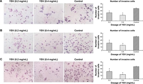
Figure 3 YSV inhibited the expression and activity of MMP-2 and MMP-9.
Notes: (A) Gelatin zymography assay for MMP-2 and MMP-9 activities from supernatant of 95D, A549, and NCI-H1299 cells after treated with YSV in vitro. Supernatants of lung cancer cells treated with different concentrations of YSV for 48 hours were collected and loaded onto 10% acrylamide (containing 0.1% gelatin) gels. After electrophoresis, the gels were washed, incubated, stained, and destained. Gelatinolytic activities were identified as clear bands on a background. The relative gelatinolytic activities of MMP-2 and MMP-9 were quantified densitometrically in relation to the activity without YSV. (B) Real-time PCR analysis of MMP-2 and MMP-9 mRNA levels in the 95D, A549, and NCI-H1299 cells after treated with YSV in vitro. Real-time PCR was performed on total RNA for detection of mRNA level of MMP-2 and MMP-9. The products were quantified using β-actin as an internal reference. (C) 2% agarose gel electrophoresis of real-time PCR production. (D) Western blot analysis of the levels of MMP-2 and MMP-9 in the 95D, A549, and NCI-H1299 cells after treated with YSV in vitro. Total proteins of lung cancer cells treated with different concentrations of YSV for 48 hours were extracted and separated by SDS-PAGE. After electrophoresis, the protein was transferred to PVDF membrane, and detection was performed with antibodies against target proteins. The products are reported as the target protein/β-actin densitometric ratio calculated by the TotalLab software to compute the relative expression of proteins. *P<0.05, compared to control group, tested using one-way ANOVA and Student–Newman–Keuls test (N=3).
Abbreviations: YSV, tyroservatide; MMP, matrix metalloproteinase; PVDF, polyvinylidene difluoride; PCR, polymerase chain reaction; SDS-PAGE, sodium dodecyl sulfate polyacrylamide gel electrophoresis; ANOVA, analysis of variance.
Abbreviations: YSV, tyroservatide; MMP, matrix metalloproteinase; PVDF, polyvinylidene difluoride; PCR, polymerase chain reaction; SDS-PAGE, sodium dodecyl sulfate polyacrylamide gel electrophoresis; ANOVA, analysis of variance.
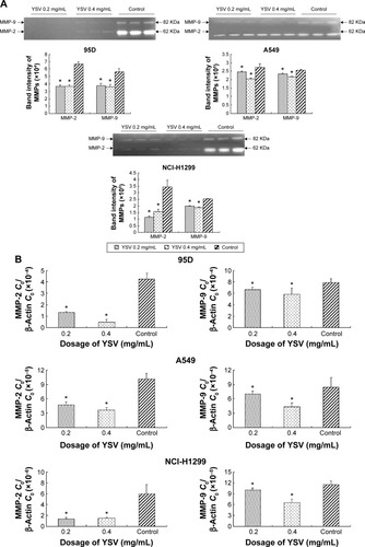
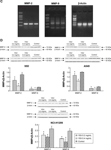
Figure 4 YSV inhibited the expression and activity of FAK in human cancer lung cells.
Notes: (A) Real-time PCR analysis of FAK mRNA level in the 95D, A549, and NCI-H1299 cells after treated with YSV in vitro. Real-time PCR was performed on total RNA for detection of FAK mRNA level. The products were quantified using β-actin as an internal reference. (B) Western blot for FAK in the 95D, A549, and NCI-H1299 cells after treated with YSV in vitro. Total proteins of 95D, A549, and NCI-H1299 cells treated with different concentrations of YSV for 48 hours were extracted and separated by SDS-PAGE. After electrophoresis, the protein was transferred to PVDF membrane, and detection was performed with antibody against target protein. The products are reported as the target protein/β-actin densitometric ratio calculated by the TotalLab software to compute the relative expression of proteins. (C) Western blot for phosphorylation of FAK Tyr397 and Tyr576/577 in the 95D, A549, and NCI-H1299 cells after treated with YSV in vitro. Total proteins of 95D, A549, and NCI-H1299 cells treated with different concentrations of YSV for 48 hours were extracted and separated by SDS-PAGE. After electrophoresis, the protein was transferred to PVDF membrane, and detection was performed with antibody against target protein. The products are reported as the target protein/β-actin densitometric ratio calculated by the TotalLab software to compute the relative expression of proteins. *P<0.05, compared to control group, tested using one-way ANOVA and Student–Newman–Keuls test (N=3).
Abbreviations: YSV, tyroservatide; FAK, focal adhesion kinase; PCR, polymerase chain reaction; SDS-PAGE, sodium dodecyl sulfate polyacrylamide gel electrophoresis; PVDF, polyvinylidene difluoride; ANOVA, analysis of variance.
Abbreviations: YSV, tyroservatide; FAK, focal adhesion kinase; PCR, polymerase chain reaction; SDS-PAGE, sodium dodecyl sulfate polyacrylamide gel electrophoresis; PVDF, polyvinylidene difluoride; ANOVA, analysis of variance.
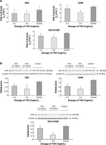
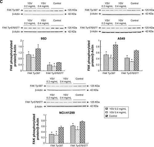
Figure 5 Real-time PCR analysis of mRNA level of integrin β1 and integrin β3 in the 95D, A549, and NCI-H1299 cells after treated with YSV in vitro.
Notes: Real-time PCR was performed on total RNA for detection of mRNA level of integrin β1 and integrin β3. The products were quantified using β-actin as an internal reference. (A) mRNA level of integrin β1 and integrin β3 in 95D cells. (B) mRNA level of integrin β1 and integrin β3 in A549 cells. (C) mRNA level of integrin β1 and integrin β3 in NCI-H1299 cells. *P<0.05, compared to control group, tested using one-way ANOVA and Student–Newman–Keuls test (N=3).
Abbreviations: PCR, polymerase chain reaction; YSV, tyroservatide; ANOVA, analysis of variance.
Abbreviations: PCR, polymerase chain reaction; YSV, tyroservatide; ANOVA, analysis of variance.
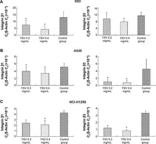
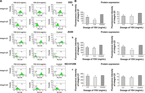
Figure 6 ECM analysis for integrin β1 (A) and integrin β3 on the membrane surface of 95D, A549, and NCI-H1299 cells (B) after treated with YSV in vitro.
Notes: 95D, A549, and NCI-H1299 cells treated with different concentrations of YSV for 48 hours were collected. Data of mean fluorescence intensity were obtained by the flow cytometer. (a) Protein expression of integrin β1 and integrin β3 in 95D cells. (b) Protein expression of integrin β1 and integrin β3 in A549 cells. (c) Protein expression of integrin β1 and integrin β3 in NCI-H1299 cells. *P<0.05, compared to control group, tested using one-way ANOVA and Student–Newman–Keuls test (N=3).
Abbreviations: ECM, extracellular matrix; YSV, tyroservatide; ANOVA, analysis of variance.
Abbreviations: ECM, extracellular matrix; YSV, tyroservatide; ANOVA, analysis of variance.