Figures & data
Table 1 In vitro growth inhibitory activity of BS-181 on gastric cancer and normal cell lines
Figure 1 BS-181 decreased the migration and invasion ability of BGC823 cells.
Notes: BGC823 cells were treated with BS-181 for 24 hours at indicated concentrations. Migration and invasion assay of BGC823 cells (A). BS-181 significantly decreased the number of migrated (B) and invaded (C) cells. Compared with control group *P<0.05 and **P<0.01; compared to other groups ##P<0.01.
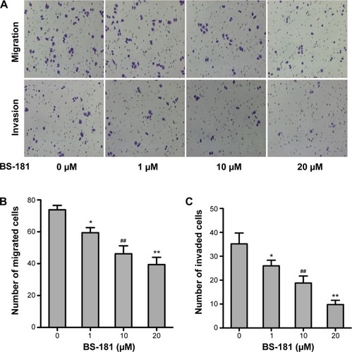
Figure 2 BS-181 induced cell apoptosis (A) and regulated apoptosis-related protein expressions (B and C).
Notes: BS-181-induced BGC823 cell apoptosis in a dose- and time-dependent manner. Additionally, BS-181 increased expression of proapoptotic proteins and decreased expression of antiapoptotic proteins. Compared with control group *P<0.05 and **P<0.01; compared to other groups #P<0.05 and ##P<0.01.
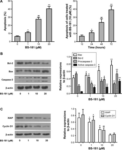
Figure 3 BS-181-impaired cell cycle progression in BGC823 cells.
Notes: Cell cycle distribution was analyzed by flow cytometry (A). Data are presented as the mean of triplicate experiments (B). Administration of BS-181 significantly increased the percentage of cells in the G0/G1 phase, and significantly decreased the S and G2/M phase fractions.
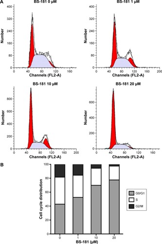
Table 2 Inhibition of cyclin-dependent protein kinase activity by BS-181
Figure 4 BS-181-inhibited phosphorylation of CDK7 substrates.
Notes: Whole cell lysates were prepared from BGC823 cells treated with BS-181 for 4 hours at indicated concentrations. Immunoblotting was carried out using antibodies for RNA polymerase II or Pol II phosphorylated at Ser5 in the C-terminal domain (A). Data are presented as mean ± SD of three independent experiments (B). Compared to other groups #P<0.05 and ##P<0.01.
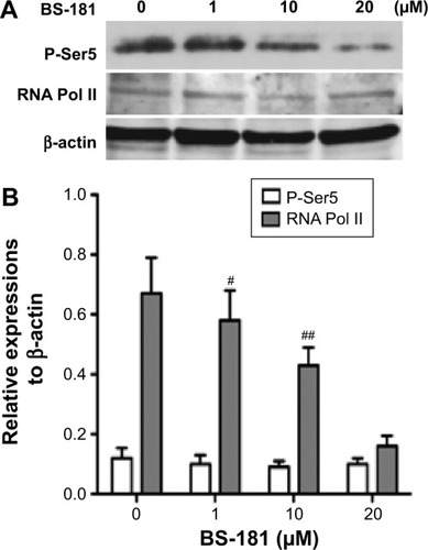
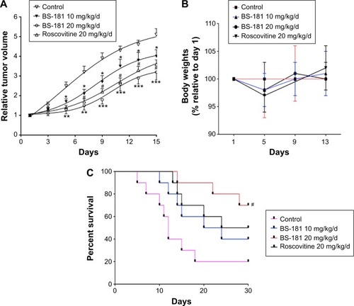
Figure 5 BS-181-inhibited gastric cancer growth in vivo and increased survival rate.
Notes: The change in tumor volume (A) was determined for each animal, as tumor volume relative to the tumor volume of each animal at day 1; mice body weights (B) were similar between groups for a 14-day observation; treatment with BS-181 significantly increased survival rate in an additional 30-day observation (C); roscovitine was used as a positive control. Compared to control group *P<0.05, **P<0.01, and ***P<0.001. Compared to other groups #P<0.05.