Figures & data
Table 1 Determination of doxorubicin IC50 in different tumor cell lines
Figure 1 Different doxorubicin sensitivity and topoisomerase IIβ binding protein 1 (TopBP1) expression in lung cancer cells.
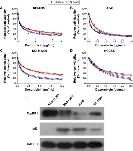
Figure 2 Measurement of cell proliferation in lung cancer cells.
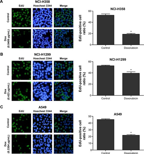
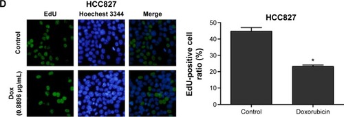
Figure 3 TopBP1 was involved in the chemoresistance of tumor cells.
Abbreviations: TopBP1, topoisomerase IIβ binding protein 1; siRNA, small interfering RNA.
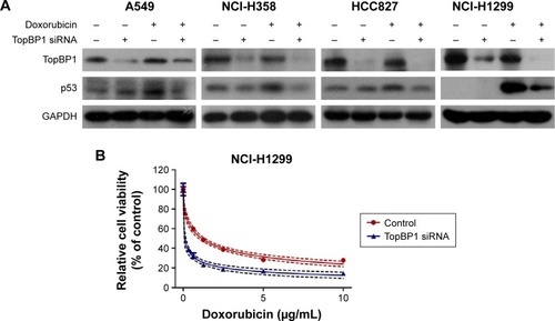
Figure 4 Measurement of cell proliferation in TopBP1 siRNA-transfected lung cancer cells.
Abbreviations: TopBP1, topoisomerase IIβ binding protein 1; siRNA, small interfering RNA.
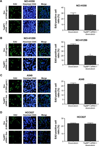
Figure 5 Upregulation of p53 by topoisomerase IIβ binding protein 1 (TopBP1) in NCI-H1299 cells.
Abbreviations: Ctrl, control; Dox, doxorubicin; TopBP1, topoisomerase IIβ binding protein 1.
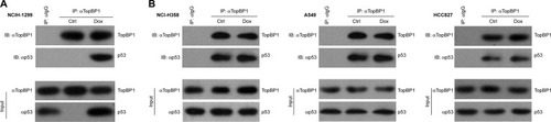
Figure S1 Cell viability (A) and apoptosis (B) in doxorubicin (Dox)-treated non-small cell lung cancer cells before and after p53 knockdown. * represents statistical significance with P<0.05.
Figure S2 Effect of TopBP1 in non-small cell lung cancer cells.