Figures & data
Figure 1 Study design (in vivo).
Abbreviations: MGL, magnesium isoglycyrrhizinate; ALT, alanine aminotransferase; AST, aspartate aminotransferase; H&E, hematoxylin and eosin; Con A, concanavalin A; PBMC, peripheral blood mononuclear cell.
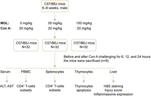
Figure 2 Liver function tests and injury scoring.
Notes: (A) Injury score of liver injury. (B) Serum ALT levels. (C) Serum AST levels. The data on the graphs indicate P values (treated group vs control group).
Abbreviations: ALT, alanine aminotransferase; Con A, concanavalin A; AST, aspartate aminotransferase; MGL, magnesium isoglycyrrhizinate.
Abbreviations: ALT, alanine aminotransferase; Con A, concanavalin A; AST, aspartate aminotransferase; MGL, magnesium isoglycyrrhizinate.
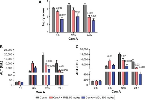
Figure 3 H&E staining of liver tissue.
Notes: MGL administration significantly improved liver injury compared with merely challenging with Con A. The blue arrow indicates the infiltrating inflammatory cells in necrotic area (200×).
Abbreviations: Con A, concanavalin A; MGL, magnesium isoglycyrrhizinate; H&E, hematoxylin and eosin. subsets and ALT levels before and after Con A challenging
Abbreviations: Con A, concanavalin A; MGL, magnesium isoglycyrrhizinate; H&E, hematoxylin and eosin. subsets and ALT levels before and after Con A challenging
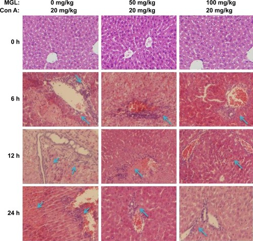
Table 1 The relationship between percentage of CD4+ T-cells subsets and ALT levels before and after Con Achallenging
Figure 4 The subsets of CD4+ T-cells in PBMCs.
Notes: (A) Shows the frequency analysis of CD4+CD25−CD69+ T-cells, representing results from control mice. (B) The frequency of CD4+CD25−CD69+ T-cells in PBMCs, before and after Con A challenge for 6, 12, and 24 hours. (C) The frequency of CD4+CD25+CD69+ T-cells in PBMCs, before and after Con A challenge for 6, 12, and 24 hours. The data on the graphs indicate P values (treated group vs control group).
Abbreviations: Con A, concanavalin A; MGL, magnesium isoglycyrrhizinate; PBMC, peripheral blood mononuclear cell; SSC, side scatter; FSC, forward scatter.
Abbreviations: Con A, concanavalin A; MGL, magnesium isoglycyrrhizinate; PBMC, peripheral blood mononuclear cell; SSC, side scatter; FSC, forward scatter.
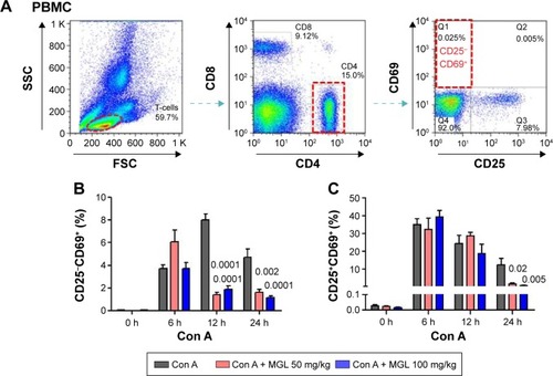
Figure 5 The subsets of CD4+ T-cells in splenocytes.
Notes: (A) Shows the frequency analysis of CD4+CD25−CD69+ T-cells, representing results from control mice. (B) The frequency of CD4+CD25−CD69+ T-cells in splenocytes, before and after Con A challenge for 6, 12, and 24 hours. (C) The frequency of CD4+CD25+CD69+ T-cells in splenocytes, before and after Con A challenge for 6, 12, and 24 hours. The data on the graphs indicate P values (treated group vs control group).
Abbreviations: Con A, concanavalin A; MGL, magnesium isoglycyrrhizinate; SSC, side scatter; FSC, forward scatter.
Abbreviations: Con A, concanavalin A; MGL, magnesium isoglycyrrhizinate; SSC, side scatter; FSC, forward scatter.
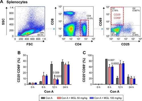
Figure 6 The apoptosis of T-cell subsets in thymocytes.
Notes: (A) Shows the analyzing method of CD4+ T-cell apoptosis in thymus. (B) CD4+ T-cell apoptosis in thymocytes, before and after Con A challenge for 6, 12, and 24 hours. (C) CD8+ T-cell apoptosis in thymocytes, before and after Con A challenge for 6, 12, and 24 hours. The data on the graphs indicate P values (treated group vs control group).
Abbreviations: Con A, concanavalin A; MGL, magnesium isoglycyrrhizinate; SSC, side scatter; FSC, forward scatter.
Abbreviations: Con A, concanavalin A; MGL, magnesium isoglycyrrhizinate; SSC, side scatter; FSC, forward scatter.
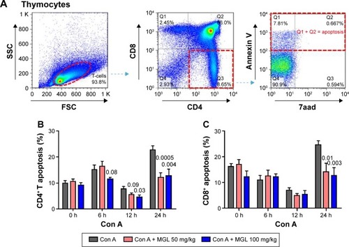
Figure 7 The proliferation of cells in the CD4+ T-cell subset before and after MGL administration and/or Con A challenging.
Notes: The upper part of figure shows the study design in vitro. (A) The CD4+ T-cells were isolated from splenocytes using a specific CD4+ T-cells isolation kit and were then cultured in plates. (B) Con A challenging significantly increased the CD25−CD69+ subset of CD4+ T-cells, but the 5 mg/mL MGL almost completely eliminated proliferation of the CD25−CD69+ subset of cells after Con A challenge for 6, 12, and 24 hours. (C) Con A challenging significantly increased the CD25+CD69+ subset of CD4+ T-cells, but the 5 mg/mL MGL almost completely eliminated proliferation of the CD25+CD69+ subset of cells after Con A challenge for 6, 12, and 24 hours. The data on the graphs indicate P values (treated group vs control group).
Abbreviations: Ab, antibody; Con A, concanavalin A; MGL, magnesium isoglycyrrhizinate; SSC, side scatter; FSC, forward scatter.
Abbreviations: Ab, antibody; Con A, concanavalin A; MGL, magnesium isoglycyrrhizinate; SSC, side scatter; FSC, forward scatter.
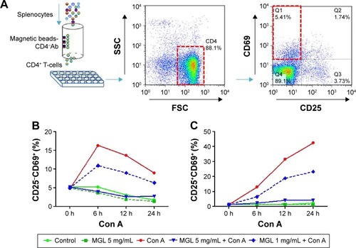
Figure 8 The inflammasome expression in liver tissue.
Notes: After Con A challenge for 6, 12, and 24 hours, the NALP3, NLRP6, and caspase-3 expression increased. (A) Shows the results of the Western blotting analysis, and (B) shows the plots of pixel intensity. For simplicity, representative blots of actin and GAPDH are shown. MGL administration significantly downregulated inflammasome expression, in which caspase-3 decreased in a dose-dependent manner.
Abbreviations: Con A, concanavalin A; MGL, magnesium isoglycyrrhizinate.
Abbreviations: Con A, concanavalin A; MGL, magnesium isoglycyrrhizinate.
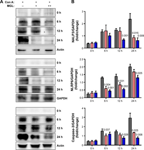
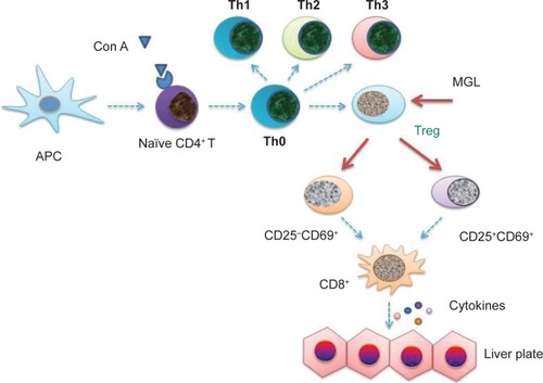
Figure 9 The possible target in the pathogensis of autoimmune hepatitis.
Notes: The blue arrow indicates that the possible molecular mechanism of pathogenesis of autoimmune hepatitis. The red arrows indicate the possible targets of MGL’s proliferation inhibiting-capacity of the regulatory T-cell subsets. Based on these data, MGL inhibiting the expression of inflammasome may be a downstream event in CD4+ T-cell subsets regulation.
Abbreviations: APC, antigen presenting cell; MGL, magnesium isoglycyrrhizinate; Con A, concanavalin A; Treg, T regulator cell.
Abbreviations: APC, antigen presenting cell; MGL, magnesium isoglycyrrhizinate; Con A, concanavalin A; Treg, T regulator cell.