Figures & data
Figure 1 Effects of intrastriatal administration of Tat-fusion interfering peptide (Tat-D1Ri) on D1R–GluN1 interactions.
Notes: Tat-D1Ri, Tat-D1Rc, or saline were locally injected into the striatum of the normal adult rat at a rate of 0.2 µL/min. Rat striatal tissues were then used for coimmunoprecipitation experiments to validate the efficacy and selectivity of interfering peptide. The intrastriatal injection of Tat-D1Ri rather than Tat-D1Rc caused reduction of D1R–GluN1 interactions, which demonstrated the effectiveness of Tat-D1Ri.
Abbreviations: Tat-D1Rc, Tat-fusion control peptide; Ig, Immunoglobulin; IP, immunoprecipitation; IB, immunoblot.
Abbreviations: Tat-D1Rc, Tat-fusion control peptide; Ig, Immunoglobulin; IP, immunoprecipitation; IB, immunoblot.
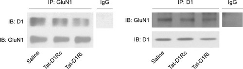
Figure 2 Effects of intrastriatal administration of Tat-D1Ri on established dyskinetic behaviors in 6-OHDA-lesioned rats.
Notes: LID rat model received intrastriatal administration of Tat-D1Ri, Tat-D1Rc, or saline 4 hours before levodopa on day 22. ALO AIM scores were compared after l-dopa injection at days 21 and 22. Data are expressed as means ± SEM. Tat-D1Ri alleviated the dyskinetic behavioral manifested by the reduction of ALO AIM scores. *P=0.05 versus vehicle plus l-dopa.
Abbreviations: 6-OHDA, 6-hydroxydopamine; l-dopa, L-3,4-dihydroxyphenylalanine; LID, l-dopa-induced dyskinesia; SEM, standard error of mean; Tat-D1Ri, Tat-fusion interfering peptide; Tat-D1Rc, Tat-fusion control peptide; ALO AIM, axial, limb, and orolingual movements.
Abbreviations: 6-OHDA, 6-hydroxydopamine; l-dopa, L-3,4-dihydroxyphenylalanine; LID, l-dopa-induced dyskinesia; SEM, standard error of mean; Tat-D1Ri, Tat-fusion interfering peptide; Tat-D1Rc, Tat-fusion control peptide; ALO AIM, axial, limb, and orolingual movements.
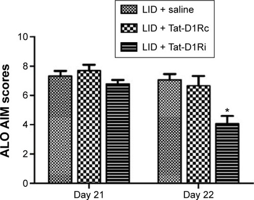
Figure 3 Effects of intrastriatal administration of Tat-D1Ri on DARPP-32 phosphorylation level after levodopa treatment in 6-OHDA-lesioned rats.
Notes: Representative Western blots are shown above the quantification of immunoblot results. Note that Tat-D1Ri rather than Tat-D1Rc substantially blocked l-dopa-stimulated phosphorylation of DARPP-32 at Thr34. Data are expressed as means ± SEM. *P=0.05 versus PD control; #P=0.05 versus saline plus l-dopa.
Abbreviations: 6-OHDA, 6-hydroxydopamine; DARPP-32, dopamine- and cAMP-regulated phosphoprotein of 32 kDa; GAPDH, glyceraldehyde 3-phosphate dehydrogenase; l-dopa, L-3,4-dihydroxyphenylalanine; LID, l-dopa-induced dyskinesia; P-DARPP-32, phosphorylated DARPP-32; SEM, standard error of mean; Tat-D1Ri, Tat-fusion interfering peptide; Tat-D1Rc, Tat-fusion control peptide; PD, Parkinson’s disease.
Abbreviations: 6-OHDA, 6-hydroxydopamine; DARPP-32, dopamine- and cAMP-regulated phosphoprotein of 32 kDa; GAPDH, glyceraldehyde 3-phosphate dehydrogenase; l-dopa, L-3,4-dihydroxyphenylalanine; LID, l-dopa-induced dyskinesia; P-DARPP-32, phosphorylated DARPP-32; SEM, standard error of mean; Tat-D1Ri, Tat-fusion interfering peptide; Tat-D1Rc, Tat-fusion control peptide; PD, Parkinson’s disease.
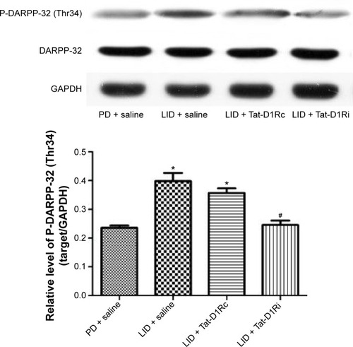
Figure 4 Effects of intrastriatal administration of Tat-D1Ri on membrane NMDA subunit expression.
Notes: Protein levels were evaluated by Western blotting of proteins extracted from the lesioned striatum of the rat brains. They were assessed in extracts from 6-OHDA-lesioned rats treated with vehicle, pulsatile l-dopa plus intrastriatal administration of saline, pulsatile l-dopa plus intrastriatal administration of Tat-D1Rc or Tat-D1Ri. (A, B) Representative Western blot analysis and densitometric analysis of GluN1 in the membrane fraction; (C, D) representative Western blot analysis and densitometric analysis of GluN2A in the membrane fraction; (E, F) representative Western blot analysis and densitometric analysis of GluN2A in the membrane fraction. Data are expressed as means ± SEM. *P=0.05 versus saline plus l-dopa.
Abbreviations: 6-OHDA, 6-hydroxydopamine; l-dopa, L-3,4-dihydroxyphenylalanine; GAPDH, glyceraldehyde 3-phosphate dehydrogenase; LID, l-dopa-induced dyskinesia; SEM, standard error of mean; Tat-D1Ri, Tat-fusion interfering peptide; Tat-D1Rc, Tat-fusion control peptide; NMDA, N-methyl-d-aspartate; PD, Parkinson’s disease.
Abbreviations: 6-OHDA, 6-hydroxydopamine; l-dopa, L-3,4-dihydroxyphenylalanine; GAPDH, glyceraldehyde 3-phosphate dehydrogenase; LID, l-dopa-induced dyskinesia; SEM, standard error of mean; Tat-D1Ri, Tat-fusion interfering peptide; Tat-D1Rc, Tat-fusion control peptide; NMDA, N-methyl-d-aspartate; PD, Parkinson’s disease.
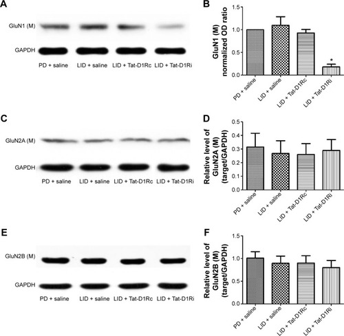
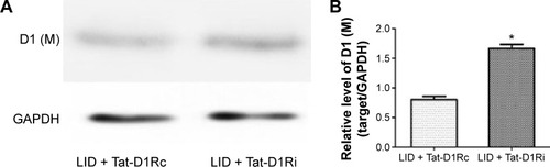
Figure 5 Effects of intrastriatal administration of Tat-D1Ri on membrane D1R subunit expression.
Notes: Protein levels were evaluated by Western blotting of proteins extracted from the lesioned striatum of the rat brains. They were assessed in extracts from 6-OHDA-lesioned rats treated with pulsatile l-dopa plus intrastriatal administration of Tat-D1Rc or Tat-D1Ri. (A) Representative Western blot analysis of D1R in the membrane fraction; (B) densitometric analysis of two blots with specific protein signals normalized to the corresponding GAPDH levels. Data are expressed as means ± SEM. Statistical analysis was conducted by independent-samples t-test. *P=0.05 versus LID + Tat-D1Rc.
Abbreviations: 6-OHDA, 6-hydroxydopamine; l-dopa, L-3,4-dihydroxyphenylalanine; LID, l-dopa-induced dyskinesia; SEM, standard error of mean; Tat-D1Ri, Tat-fusion interfering peptide; Tat-D1Rc, Tat-fusion control peptide; GAPDH, glyceraldehyde 3-phosphate dehydrogenase.
Abbreviations: 6-OHDA, 6-hydroxydopamine; l-dopa, L-3,4-dihydroxyphenylalanine; LID, l-dopa-induced dyskinesia; SEM, standard error of mean; Tat-D1Ri, Tat-fusion interfering peptide; Tat-D1Rc, Tat-fusion control peptide; GAPDH, glyceraldehyde 3-phosphate dehydrogenase.